

Tubulin Folding Cofactor TBCB is a Target of the Salmonella Effector Protein SseK1
- PMID: 32366039
- PMCID: PMC7246435
- DOI: 10.3390/ijms21093193
Tubulin Folding Cofactor TBCB is a Target of the Salmonella Effector Protein SseK1
Abstract
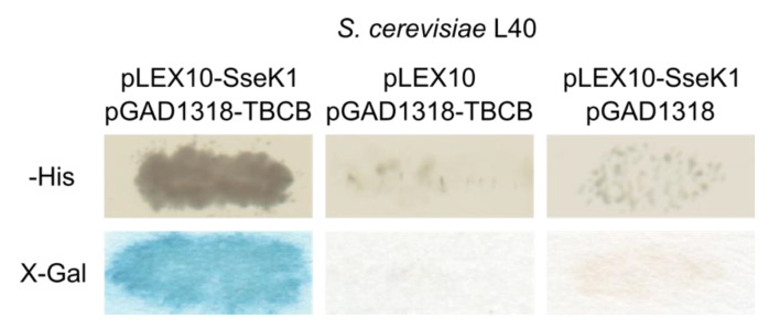
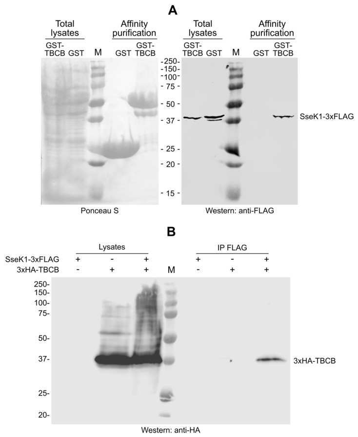
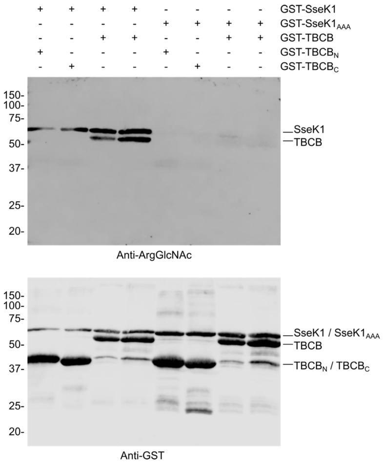
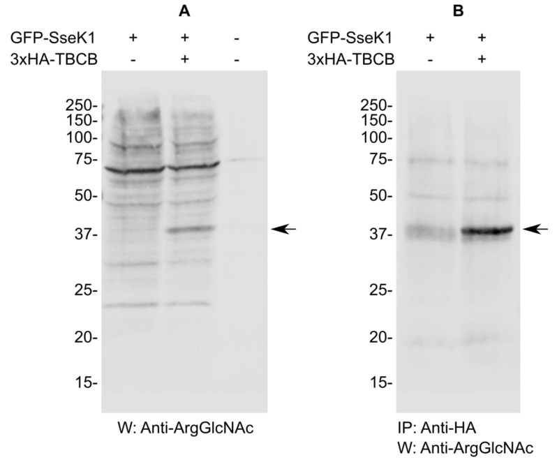
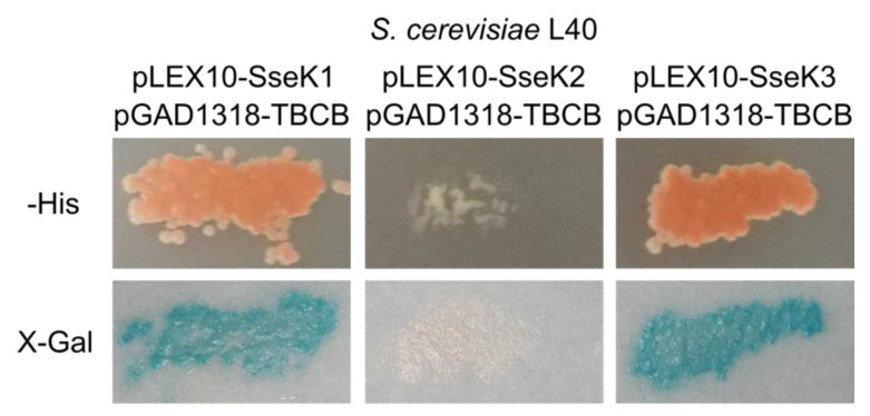
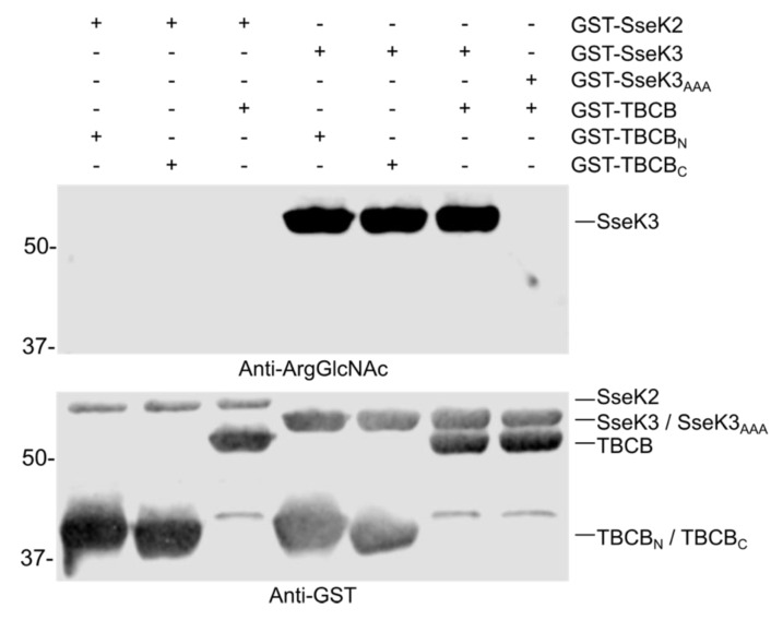
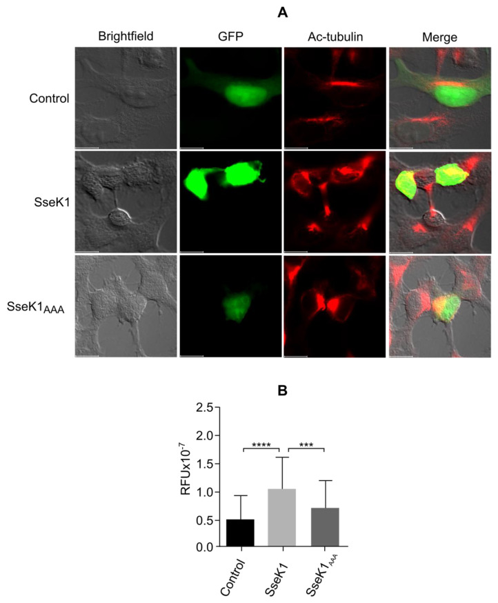
Salmonella enterica serovar Typhimurium is a human and animal pathogen that uses type III secretion system effectors to manipulate the host cell and fulfill infection. SseK1 is a Salmonella effector with glycosyltransferase activity. We carried out a yeast two-hybrid screen and have identified tubulin-binding cofactor B (TBCB) as a new binding partner for this effector. SseK1 catalyzed the addition of N-acetylglucosamine to arginine on TBCB, and its expression promoted the stabilization of the microtubule cytoskeleton of HEK293T cells. The conserved Asp-x-Asp (DxD) motif that is essential for the activity of SseK1 was required for the binding and modification of TBCB and for the effect on the cytoskeleton. Our study has identified a novel target for SseK1 and suggests that this effector may have a role in the manipulation of the host cell microtubule network to provide a safe niche for this pathogen.
Keywords: Salmonella; SseK1; TBCB; arginine N-glycosyltransferase; tubulin; type III secretion.
Conflict of interest statement
The authors declare no conflict of interest. The funders had no role in the design of the study; in the collection, analyses, or interpretation of data; in the writing of the manuscript, or in the decision to publish the work.
Figures
Similar articles
-
SseK1 and SseK3 Type III Secretion System Effectors Inhibit NF-κB Signaling and Necroptotic Cell Death in Salmonella-Infected Macrophages.Infect Immun. 2017 Feb 23;85(3):e00010-17. doi: 10.1128/IAI.00010-17. Print 2017 Mar. Infect Immun. 2017. PMID: 28069818 Free PMC article.
-
SseK1 and SseK2 are novel translocated proteins of Salmonella enterica serovar typhimurium.Infect Immun. 2004 Sep;72(9):5115-25. doi: 10.1128/IAI.72.9.5115-5125.2004. Infect Immun. 2004. PMID: 15322005 Free PMC article.
-
Effects of Salmonella enterica serovar typhimurium sseK1 on macrophage inflammation-related cytokines and glycolysis.Cytokine. 2021 Apr;140:155424. doi: 10.1016/j.cyto.2021.155424. Epub 2021 Jan 26. Cytokine. 2021. PMID: 33513526
-
Type three secretion system in Salmonella Typhimurium: the key to infection.Genes Genomics. 2020 May;42(5):495-506. doi: 10.1007/s13258-020-00918-8. Epub 2020 Feb 28. Genes Genomics. 2020. PMID: 32112371 Review.
-
[Advances in Salmonella pathogenicity island 2 type III secretion system - A review].Wei Sheng Wu Xue Bao. 2016 Apr 4;56(4):561-9. Wei Sheng Wu Xue Bao. 2016. PMID: 29717847 Review. Chinese.
Cited by
-
Arginine glycosylation regulates UDP-GlcNAc biosynthesis in Salmonella enterica.Sci Rep. 2022 Mar 28;12(1):5293. doi: 10.1038/s41598-022-09276-9. Sci Rep. 2022. PMID: 35351940 Free PMC article.
-
Catalytic DxD motif caged in Asx-turn and Met-aromatic interaction attenuates the pathogenic glycosylation of SseK2/NleB2 effectors.Sci Rep. 2022 Nov 11;12(1):19288. doi: 10.1038/s41598-022-22803-y. Sci Rep. 2022. PMID: 36369343 Free PMC article.
-
Host-Pathogen Interaction 3.0.Int J Mol Sci. 2022 Oct 24;23(21):12811. doi: 10.3390/ijms232112811. Int J Mol Sci. 2022. PMID: 36361600 Free PMC article.
-
Salmonella T3SS effector SseK1 arginine-glycosylates the two-component response regulator OmpR to alter bile salt resistance.Sci Rep. 2023 Jun 3;13(1):9018. doi: 10.1038/s41598-023-36057-9. Sci Rep. 2023. PMID: 37270573 Free PMC article.
-
Recent progress in molecular mechanisms of Salmonella effectors involved in gut epithelium invasion.Mol Biol Rep. 2025 Jun 16;52(1):601. doi: 10.1007/s11033-025-10715-9. Mol Biol Rep. 2025. PMID: 40522423 Review.
References
-
- Condry D.L.J., Nilles M.L. Introduction to Type III Secretion Systems. Methods Mol. Biol. (Clifton, N.J.) 2017;1531:1–10. - PubMed
MeSH terms
Substances
Grants and funding
LinkOut - more resources
Full Text Sources
Molecular Biology Databases
Miscellaneous
