. 2020 May 4;19(1):82.
doi: 10.1186/s12943-020-01205-6.

circNFIB1 inhibits lymphangiogenesis and lymphatic metastasis via the miR-486-5p/PIK3R1/VEGF-C axis in pancreatic cancer

Affiliations

circNFIB1 inhibits lymphangiogenesis and lymphatic metastasis via the miR-486-5p/PIK3R1/VEGF-C axis in pancreatic cancer

Yao Kong et al. Mol Cancer. .

Abstract

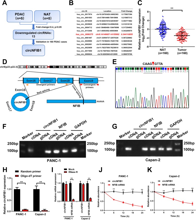
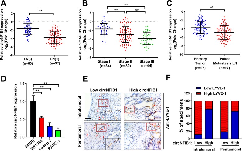
Background: Patients with lymph node (LN)-positive pancreatic ductal adenocarcinoma (PDAC) have extremely poor survival rates. Circular RNAs (circRNAs), a newly discovered type of endogenous noncoding RNAs, have been proposed to mediate the progression of diverse types of tumors. However, the role and underlying regulatory mechanisms of circRNAs in the LN metastasis of PDAC remain unknown.

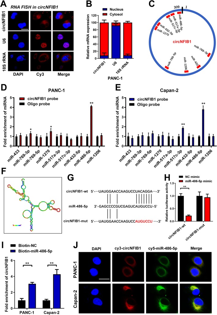
Methods: Next-generation sequencing was used to identify differentially expressed circRNAs between PDAC and normal adjacent tissues. In vitro and in vivo experiments were conducted to evaluate the functional role of circNFIB1. RNA pulldown and luciferase assays were performed to examine the binding of circNFIB1 and miR-486-5p.

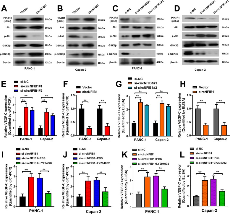
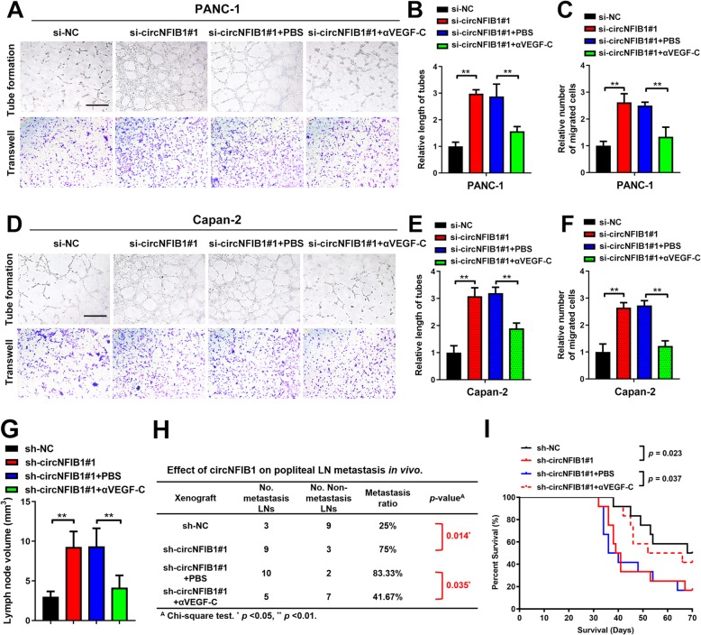
Results: In the present study, we identified that a novel circRNA (circNFIB1, hsa_circ_0086375) was downregulated in PDAC and negatively associated with LN metastasis in PDAC patients. Functionally, circNFIB1 knockdown promoted lymphangiogenesis and LN metastasis of PDAC both in vitro and in vivo. Mechanistically, circNFIB1 functioned as a sponge of miR-486-5p, and partially reversed the effect of miR-486-5p. Moreover, circNFIB1 attenuated the oncogenic effect of miR-486-5p and consequently upregulated PIK3R1 expression, which further downregulated VEGF-C expression through inhibition of the PI3K/Akt pathway, and ultimately suppressed lymphangiogenesis and LN metastasis in PDAC.

Conclusions: Our findings provide novel insight into the underlying mechanism of circRNA-mediated LN metastasis of PDAC and suggest that circNFIB1 may serve as a potential therapeutic target for LN metastasis in PDAC.

Keywords: Lymphatic metastasis; PI3K/Akt signaling pathway; PIK3R1; Pancreatic cancer; circNFIB1.


Conflict of interest statement

The authors declare no potential conflicts of interest.

Figures

Fig. 1
Fig. 1
The identification and characterization of circNFIB1 in PDAC. a Schematic illustration of the identification of circRNAs downregulated in PDAC tissues compared with NATs. b The downregulated circRNAs in PDAC identified from NGS were shown. c qRT-PCR analysis of circNFIB1 expression in PDAC tissues (n = 160) paired with NATs (n = 160). The nonparametric Mann-Whitney U test was used to compare different groups. d Schematic illustrations showed the genomic loci of circNFIB1. circNFIB1 was produced by exons 16 to 18 of NFIB. e The back-splice junction of circNFIB1 was identified by Sanger sequencing. f and g PCR analysis for circNFIB1 and NFIB in the cDNA and gDNA of PANC-1 (f) or Capan-2 (g) cells. GAPDH was used as NC. h qRT-PCR analysis of circNFIB1 expression using random primers or oligo-dT primers. i circNFIB1 and NFIB expression in PDAC cells treated with or without RNase R was assessed by qRT-PCR. j and k qRT-PCR analysis of circNFIB1 and NFIB mRNA in PANC-1 (j) or Capan-2 (k) cells treated with actinomycin D at the indicated time points. Significance level was assessed using two-tailed Student t-tests. Figures with error bars showed the standard deviations of three independent experiments. *p < 0.05 and **p < 0.01
Fig. 2
Fig. 2
CircNFIB1 is downregulated in PDAC and negatively correlates with LN metastasis. a and b circNFIB1 expression in human PDAC tissues (n = 160) according to LN status (a) and tumor stages (b) was assessed by qRT-PCR. The nonparametric Mann-Whitney U test was used to compare different groups. c qRT-PCR analyzed the expression of circNFIB1 in primary PDAC tissues and paired metastatic LNs (n = 160). The nonparametric Mann-Whitney U test was used to compare different groups. d The expression of circNFIB1 in PDAC and normal pancreatic ductal epithelial cell lines was detected by qRT-PCR. e and f Representative images (e) and percentages (f) for IHC staining showed that LYVE-1 indicated the lymphatic vessel density in PDAC tissues with differential circNFIB1 expression. Scale bars: 50 μm. Significance level was assessed using one-way ANOVA followed by Dunnett’s tests for multiple comparisons. Figures with error bars showed the standard deviations of three independent experiments. *p < 0.05 and **p < 0.01
Fig. 3
Fig. 3
CircNFIB1 suppresses lymphangiogenesis in vitro. a-d qRT-PCR analysis of circNFIB1 and NFIB expression following circNFIB1-silencing (a and b), circNFIB1 overexpression (c and d) or corresponding control PDAC cells. e-j Representative images (e and h) and histogram analysis of tube formation (f and i) and Transwell (g and j) assays by HLECs treated with conditioned medium from circNFIB1-silencing or control PDAC cells. Scale bar: 100 μm. k-p Representative images (k and n) and histogram analysis of tube formation (l and o) and Transwell (m and p) assays by HLECs treated with conditioned medium from circNFIB1-overexpressing or control PDAC cells. Scale bar: 100 μm. Significance level was assessed using two-tailed Student t-tests and one-way ANOVA followed by Dunnett’s tests for multiple comparisons. Figures with error bars showed the standard deviations of three independent experiments. *p < 0.05 and **p < 0.01
Fig. 4
Fig. 4
CircNFIB1 inhibits LN metastasis of PDAC in vivo. a and b Representative images (a) of bioluminescence and histogram analysis (b) of popliteal LN metastasis after silencing circNFIB1 (n = 12 per group). c Representative images for the nude mouse popliteal LN metastasis model. PDAC cells were injected into the footpads of the nude mice and the popliteal LNs were shown. d Representative images of enucleated popliteal LNs (n = 12 per group). e Histogram analysis of the LN volume in the indicated groups. f The ratio of popliteal LN metastasis was calculated for all groups (n = 12 per group). Significance level was assessed using two-tailed Student t-tests and one-way ANOVA followed by Dunnett’s tests for multiple comparisons. Figures with error bars showed the standard deviations of three independent experiments. *p < 0.05 and **p < 0.01
Fig. 5
Fig. 5
CircNFIB1 acts as a sponge for miR-486-5p in PDAC. a and b Representative FISH images (a) and subcellular fractionation assays (b) showed the subcellular distribution of circNFIB1 in Capan-2 cells. U6 was used as a nuclear control and 18S rRNA was used as a cytoplasmic control. Scale bar: 100 μm. c the potential target miRNAs of circNFIB1 were predicted using CircInteractome. d and e RNA pulldown assays revealed that miRNAs directly interact with circNFIB1 in PANC-1 (d) and Capan-2 (e) cells. f The secondary structure of circNFIB1 was predicted by RNAalifold. g Schematic illustrations showed the alignment of circNFIB1 with miR-486-5p and the red part indicates the mutagenesis nucleotides. h Dual luciferase reporter assays show the luciferase activity of wild-type or mutant circNFIB1 following co-transfection with either the miR-486-5p or control mimics. Relative firefly luciferase expression was normalized to that of Renilla luciferase. i RNA pulldown assays showed the RNAs captured by biotinylated miR-486-5p. j Representative FISH images showed the colocalization of circNFIB1 and miR-486-5p. Scale bar: 100 μm. Significance level was assessed using two-tailed Student t-tests and one-way ANOVA followed by Dunnett’s tests for multiple comparisons. Figures with error bars showed standard deviations of three independent experiments. *p < 0.05 and **p < 0.01
Fig. 6
Fig. 6
CircNFIB1 antagonizes miR-486-5p-mediated repression of PIK3R1 expression. a-d Representative images and a histogram analysis of tube formation and Transwell assay by HLECs treated with conditioned medium from miR-486-5p-silenced (a and b), miR-486-5p overexpressing (c and d), and corresponding control PDAC cells. Scale bar: 100 μm. e Schematic illustration showed the alignment of miR-486-5p with PIK3R1 and the red portion indicated the mutagenesis nucleotides. f Dual luciferase reporter assays showed the luciferase activity of wild type or mutant PIK3R1 following co-transfection with miR-486-5p mimic or control mimic. Relative firefly luciferase expression was normalized to that of Renilla luciferase. g and h The effect of circNFIB1-silencing or circNFIB1 overexpression on PIK3R1 expression in PDAC cells was assessed by qRT-PCR. i and j qRT-PCR analyzed the effect of circNFIB1-silencing on miR-486-5p depletion-induced PIK3R1 expression in PDAC cells. k and l Western blot analysis of circNFIB1-silencing on miR-486-5p depletion-induced PIK3R1 expression in PDAC cells. Significance level was assessed using two-tailed Student t-tests and one-way ANOVA followed by Dunnett’s tests for multiple comparisons. Figures with error bars showed the standard deviations of three independent experiments. *p < 0.05 and **p < 0.01
Fig. 7
Fig. 7
CircNFIB1 downregulates VEGF-C expression through inhibiting the PI3K/Akt signaling pathway. a-d Western blot analysis of PIK3R1, Akt, p-Akt, GSK3β, and p-GSK3β levels after the overexpression (a and b) or silencing (c and d) of circNFIB1 in PDAC cells. e and f The effect of circNFIB1-silencing (e) or circNFIB1-overexpression (f) on VEGF-C expression in PDAC was assessed by qRT-PCR. g and h ELISA analysis of VEGF-C secretion by PDAC after silencing (g) or overexpressing (h) circNFIB1. i and j The effect of LY294002 treatment on circNFIB1 depletion-induced VEGF-C expression in PDAC cells was assessed by qRT-PCR. k and l ELISA for LY294002 treatment on circNFIB1 depletion-induced VEGF-C secretion by PDAC cells. Significance level was assessed using two-tailed Student t-tests and one-way ANOVA followed by Dunnett’s tests for multiple comparisons. Figures with error bars showed the standard deviations of three independent experiments. *p < 0.05 and **p < 0.01
Fig. 8
Fig. 8
CircNFIB1 inhibits lymphangiogenesis and LN metastasis of PDAC via VEGF-C suppression. a-f Representative images (a and d) and a histogram analysis of tube formation (b and e) and Transwell (c and f) assay by HLECs treated with conditioned medium from si-NC, si-circNFIB1, si-circNFIB1 + PBS, and si-circNFIB1 + αVEGF-C PDAC cells. Scale bar: 100 μm. g Histogram analysis of the LN volume in the indicated groups. h The ratio of popliteal LN metastasis was calculated for all groups (n = 12 per group). i Kaplan-Meier survival curves for indicated groups (n = 12 per group). Significance level was assessed using two-tailed Student t-tests and one-way ANOVA followed by Dunnett’s tests for multiple comparisons. Figures with error bars showed the standard deviations of three independent experiments. *p < 0.05 and **p < 0.01
Fig. 9
Fig. 9
Clinical implication of circNFIB1 as miR-486-5p sponge in PDAC. a qRT-PCR analysis of miR-486-5p expression in PDAC tissues (n = 160) paired with NATs (n = 160). The nonparametric Mann-Whitney U test was used to compare different groups. b miR-486-5p expression in human PDAC tissues (n = 160) according to the LN status was assessed by qRT-PCR. The nonparametric Mann-Whitney U test was used to compare different groups. c and d Kaplan-Meier survival curves showed the OS (c) and DFS (d) of PDAC patients with low vs. high circNFIB1 expression. The cutoff value was the median expression of circNFIB1. e and f Kaplan-Meier survival curves showed the OS (e) and DFS (f) of PDAC patients with low vs. high miR-486-5p expression. The cutoff value was the median expression of miR-486-5p. g Correlation analysis of circNFIB1 and VEGF-C expression analyzed by qRT-PCR in PDAC tissues (n = 160). h Correlation analysis of miR-486-5p and VEGF-C expression analyzed by qRT-PCR in PDAC tissues (n = 160). i Proposed model for the role of circNFIB1 on promoting lymphangiogenesis and LN metastasis via the miR-486-5p/PIK3R1/VEGF-C axis in PDAC. *p < 0.05 and **p < 0.01

References

    1. Ellis C, Ramzy A, Kieffer TJ. Regenerative medicine and cell-based approaches to restore pancreatic function. Nat Rev Gastroenterol Hepatol. 2017;14:612–628. doi: 10.1038/nrgastro.2017.93. - DOI - PubMed
    1. Makohon-Moore A, Iacobuzio-Donahue CA. Pancreatic cancer biology and genetics from an evolutionary perspective. Nat Rev Cancer. 2016;16:553–565. doi: 10.1038/nrc.2016.66. - DOI - PMC - PubMed
    1. Vincent A, Herman J, Schulick R, Hruban RH, Goggins M. Pancreatic cancer. Lancet. 2011;378:607–620. doi: 10.1016/S0140-6736(10)62307-0. - DOI - PMC - PubMed
    1. Philip PA, Mooney M, Jaffe D, Eckhardt G, Moore M, Meropol N, Emens L, O'Reilly E, Korc M, Ellis L, et al. Consensus report of the national cancer institute clinical trials planning meeting on pancreas cancer treatment. J Clin Oncol. 2009;27:5660–5669. doi: 10.1200/JCO.2009.21.9022. - DOI - PMC - PubMed
    1. Groot VP, Gemenetzis G, Blair AB, Rivero-Soto RJ, Yu J, Javed AA, Burkhart RA, Rinkes I, Molenaar IQ, Cameron JL, et al. Defining and predicting early recurrence in 957 patients with resected pancreatic ductal adenocarcinoma. Ann Surg. 2019;269:1154–1162. doi: 10.1097/SLA.0000000000002734. - DOI - PMC - PubMed

Publication types

MeSH terms