Review
. 2020 Jun 19;295(25):8425-8441.
doi: 10.1074/jbc.REV120.009309. Epub 2020 May 4.

Malaria parasite plasmepsins: More than just plain old degradative pepsins

Affiliations
Review

Malaria parasite plasmepsins: More than just plain old degradative pepsins

Armiyaw S Nasamu et al. J Biol Chem. .

Abstract

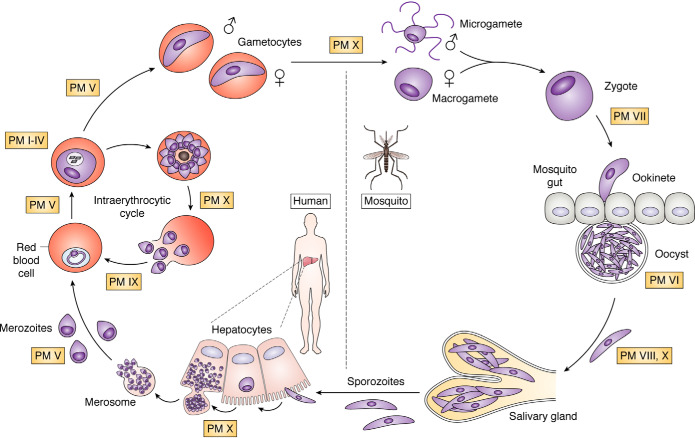
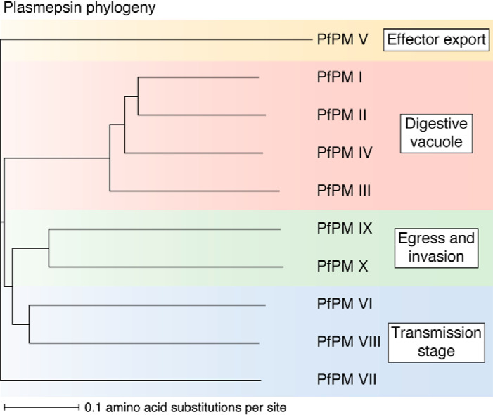
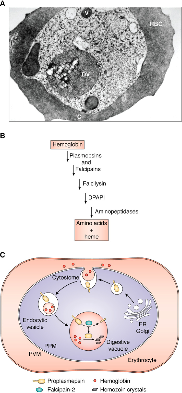
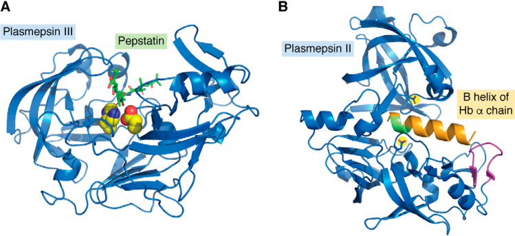
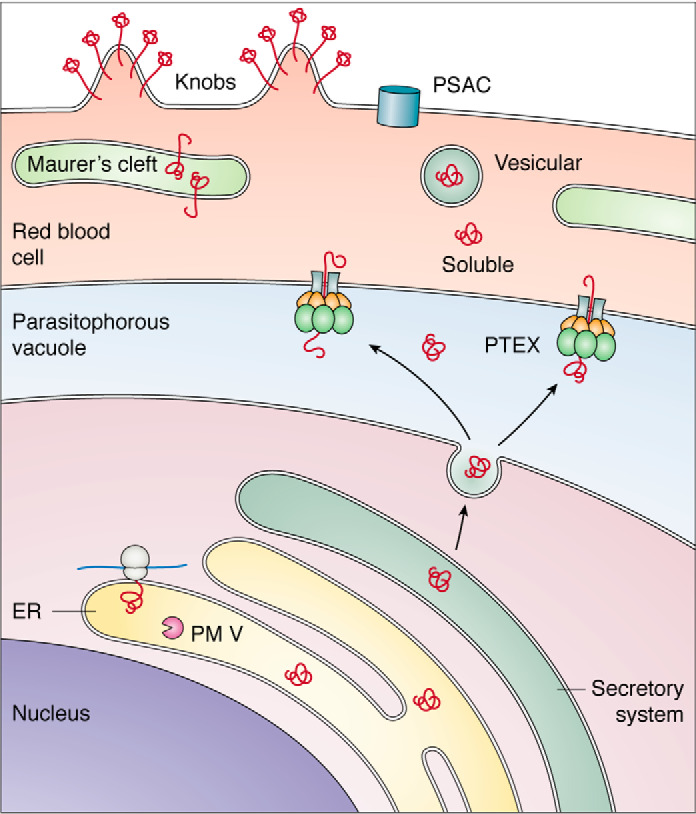
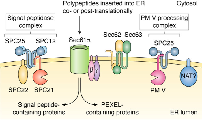
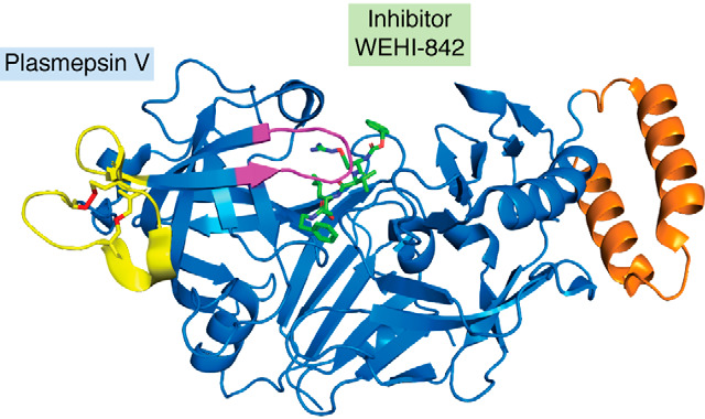
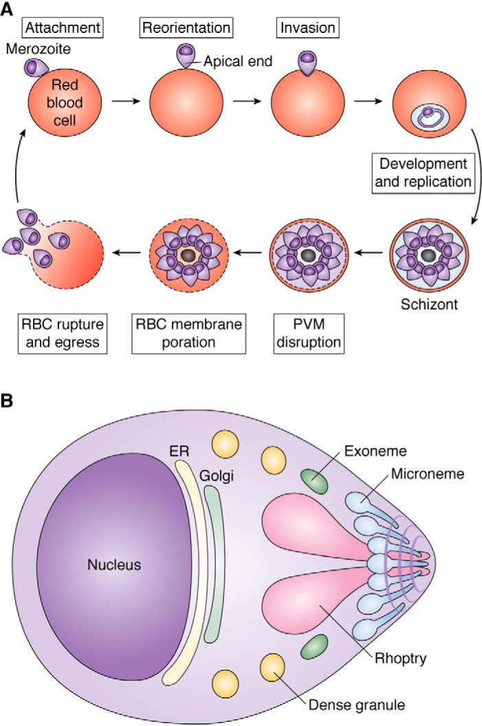
Plasmepsins are a group of diverse aspartic proteases in the malaria parasite Plasmodium Their functions are strikingly multifaceted, ranging from hemoglobin degradation to secretory organelle protein processing for egress, invasion, and effector export. Some, particularly the digestive vacuole plasmepsins, have been extensively characterized, whereas others, such as the transmission-stage plasmepsins, are minimally understood. Some (e.g. plasmepsin V) have exquisite cleavage sequence specificity; others are fairly promiscuous. Some have canonical pepsin-like aspartic protease features, whereas others have unusual attributes, including the nepenthesin loop of plasmepsin V and a histidine in place of a catalytic aspartate in plasmepsin III. We have learned much about the functioning of these enzymes, but more remains to be discovered about their cellular roles and even their mechanisms of action. Their importance in many key aspects of parasite biology makes them intriguing targets for antimalarial chemotherapy. Further consideration of their characteristics suggests that some are more viable drug targets than others. Indeed, inhibitors of invasion and egress offer hope for a desperately needed new drug to combat this nefarious organism.

Keywords: antimalarial chemotherapy; aspartic protease; digestive vacuole; hemoglobin; malaria; maturase; parasitology; plasmodium; protease; protozoan; transmission.


Conflict of interest statement

Conflict of interest—The authors declare that they have no conflicts of interest with the contents of this article.

Figures

Figure 1.
Figure 1.
Life cycle of the malaria parasite. Sporozoites from the salivary glands of an infected mosquito (bottom) make their way to the liver, infect hepatocytes, replicate to thousands of infective merozoites, and bud off as merosomes that rupture into the bloodstream. The merozoites invade RBCs, replicate, and multiply in the intraerythrocytic cycle. Some differentiate into male and female gametocytes that are taken up into the mosquito, where they develop. In the mosquito midgut, parasites egress from the RBCs as gametes, mate to form zygotes, and differentiate into ookinetes that traverse the midgut and become oocysts. They replicate and differentiate into sporozoites that migrate to the salivary glands, where they are ready for transmission to the next human upon mosquito bite. Points in the life cycle at which plasmepsins are thought to function are labeled.
Figure 2.
Figure 2.
Plasmepsin phylogeny. Sequences for PMs I–X were obtained from PlasmoDB (release 46), aligned using MUSCLE (“Multiple Sequence Comparison by Log-Expectation”, EMBL) (189), and visualized using iTOL (Interactive Tree of Life) (190).
Figure 3.
Figure 3.
Hemoglobin ingestion and digestion. A, electron micrograph of a P. falciparum-infected erythrocyte. C, cytostome; V, vesicle; DV, digestive vacuole. Adapted from Ref. . This research was originally published in Proceedings of the National Academy of Sciences of the United States of America. Goldberg, D. E., Slater, A. F. G., Cerami, A., and Henderson, G. B. Hemoglobin degradation in the malaria parasite Plasmodium falciparum: an ordered process in a unique organelle. Proc. Natl. Acad. Sci. U.S.A. 1990; 87:2931–2935. © United States National Academy of Sciences. B, semi-ordered pathway of hemoglobin degradation. Plasmepsins and falcipains are involved in the initial steps of catabolism and are partially redundant. Falcilysin recognizes oligopeptides. DPAP1 cleaves two residues off of the N terminus of hemoglobin fragments. Aminopeptidases finish the digestion, resulting in free amino acids. Heme is liberated during the initial steps of proteolysis; most is sequestered in the digestive vacuole as hemozoin. C, plasmepsin targeting pathway follows the hemoglobin internalization route. Plasmepsins are made in the ER and traverse the secretory system as type II integral membrane protein precursors (ball and stick). They traffic to the cytostome, a hemoglobin ingestion apparatus that spans the two membranes at the parasite surface. They are internalized with their substrate cargo (hemoglobin) and are delivered to the digestive vacuole surface, where they are cleaved by falcipain-2. PPM, parasite plasma membrane. Black bars, hemozoin crystals that accumulate after heme release.
Figure 4.
Figure 4.
A, crystal structure of PM III complexed with pepstatin. Shown is a ribbon structure (blue) with Asp215 and His32 highlighted in yellow with red and blue heteroatoms. Pepstatin is in green with red oxygens. The figure was constructed from PDB entry 3FNT. B, crystal structure of PM II (blue) with the B helix of the hemoglobin α chain modeled in orange. The α33–34 cleavage site is green; the helix-interacting loop is magenta; from PDB entry 1PSE. Created using PyMOL Molecular Graphics System, Version 2.3.
Figure 5.
Figure 5.
Biosynthesis and trafficking of exported proteins. Proteins are synthesized in the ER and traverse the secretory system (green) to the parasitophorous vacuole (blue), where they are recognized by the PTEX translocon and exported into the RBC (pink). In the RBC, effectors can be soluble, can reside in the Golgi-like Maurer's clefts that are established by the parasite in the host cell, can be vesicular, or can go to the RBC surface, forming nutrient acquisition channels (PSAC) or clustering variant surface cytoadhesins in knobs at the RBC surface.
Figure 6.
Figure 6.
Model for signal processing in the ER. Secretory proteins are cleaved immediately after translation either by the canonical signal peptidase (SPC21) complex (left) or a noncanonical PM V–processing complex (right). NAT, putative N-acetyltransferase.
Figure 7.
Figure 7.
Crystal structure of PM V with the inhibitor WEHI-842 bound (green). Highlighted are the flap over the active site (magenta), nepenthesin loop (yellow, with Cys-Cys bonds in red), and helix-turn-helix (orange); from PDB entry 4ZL4.
Figure 8.
Figure 8.
Plasmodium invasion and egress. A, an infective merozoite (top left) attaches to an RBC, reorients so that its apical end is in contact with the host cell, and invades. It develops, replicates, and divides, forming a schizont (bottom right). When ready to egress, it disrupts the PVM and then the RBC membrane, and finally merozoites rupture out of the RBC explosively (bottom left). B, schematic of a merozoite, showing secretory organelles whose function is influenced by plasmepsins. Exonemes discharge to initiate egress. Then micronemes discharge to secrete RBC adhesion ligands onto the cell surface. Next, rhoptries discharge to prepare the parasite and host cell for invasion and to condition the PVM. Finally, dense granules discharge to effect maturation of the parasitophorous vacuole.

References

    1. Goldberg D. E., Slater A. F. G., Beavis R., Chait B., Cerami A., and Henderson G. B. (1991) Hemoglobin degradation in the human malaria pathogen Plasmodium falciparum: a catabolic pathway initiated by a specific aspartic protease. J. Exp. Med. 173, 961–969 10.1084/jem.173.4.961 - DOI - PMC - PubMed
    1. Francis S. E., Gluzman I. Y., Oksman A., Knickerbocker A., Mueller R., Bryant M. L., Sherman D. R., Russell D. G., and Goldberg D. E. (1994) Molecular characterization and inhibition of a Plasmodium falciparum aspartic hemoglobinase. EMBO J. 13, 306–317 10.1002/j.1460-2075.1994.tb06263.x - DOI - PMC - PubMed
    1. Dame J. B., Reddy G. R., Yowell C. A., Dunn B. M., Kay J., and Berry C. (1994) Sequence, expression and modeled structure of an aspartic proteinase from the human malaria parasite Plasmodium falciparum. Mol. Biochem. Parasitol. 64, 177–190 10.1016/0166-6851(94)90024-8 - DOI - PubMed
    1. Dame J. B., Yowell C. A., Omara-Opyene L., Carlton J. M., Cooper R. A., and Li T. (2003) Plasmepsin 4, the food vacuole aspartic proteinase found in all Plasmodium spp. infecting man. Mol. Biochem. Parasitol. 130, 1–12 10.1016/S0166-6851(03)00137-3 - DOI - PubMed
    1. Banerjee R., Liu J., Beatty W., Pelosof L., Klemba M., and Goldberg D. E. (2002) Four plasmepsins are active in the Plasmodium falciparum food vacuole, including a protease with an active-site histidine. Proc. Natl. Acad. Sci. U.S.A. 99, 990–995 10.1073/pnas.022630099 - DOI - PMC - PubMed

Publication types

MeSH terms

LinkOut - more resources