. 2020 Jun 4;55(6):2000544.
doi: 10.1183/13993003.00544-2020. Print 2020 Jun.

Nosocomial outbreak of COVID-19 pneumonia in Wuhan, China

Affiliations

Nosocomial outbreak of COVID-19 pneumonia in Wuhan, China

Xiaorong Wang et al. Eur Respir J. .

Abstract

Background: The novel coronavirus, severe acute respiratory syndrome coronavirus 2 (SARS-CoV-2), infected over 3300 healthcare workers in early 2020 in China. Little information is known about nosocomial infections of healthcare workers in the initial period. We analysed data from healthcare workers with nosocomial infections in Wuhan Union Hospital (Wuhan, China) and their family members.

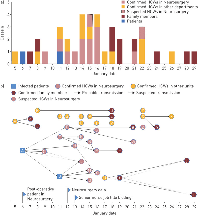
Methods: We collected and analysed data on exposure history, illness timelines and epidemiological characteristics from 25 healthcare workers with laboratory-confirmed coronavirus disease 2019 (COVID-19) and two healthcare workers in whom COVID-19 was highly suspected, as well as 10 of their family members with COVID-19, between 5 January and 12 February 2020. The demographics and clinical features of the 35 laboratory-confirmed cases were investigated and viral RNA of 12 cases was sequenced and analysed.

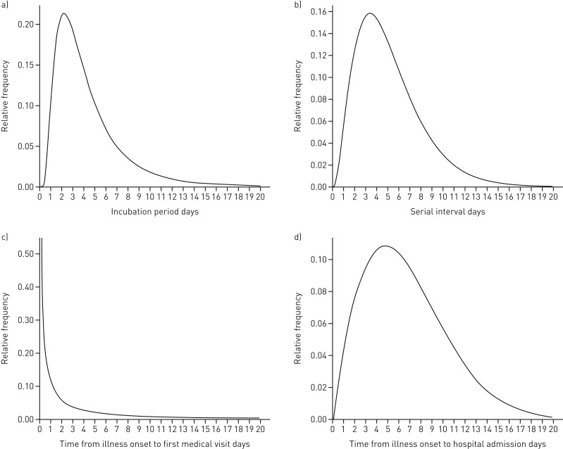
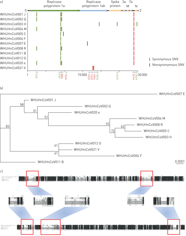
Results: Nine clusters were found among the patients. All patients showed mild to moderate clinical manifestation and recovered without deterioration. The mean period of incubation was 4.5 days, the mean±sd clinical onset serial interval (COSI) was 5.2±3.2 days, and the median virus shedding time was 18.5 days. Complete genomic sequences of 12 different coronavirus strains demonstrated that the viral structure, with small irrelevant mutations, was stable in the transmission chains and showed remarkable traits of infectious traceability.

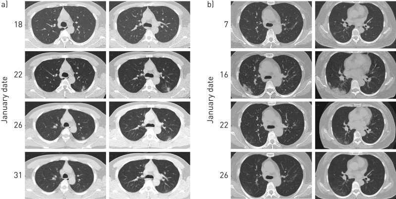
Conclusions: SARS-CoV-2 can be rapidly transmitted from person to person, regardless of whether they have symptoms, in both hospital settings and social activities, based on the short period of incubation and COSI. The public health service should take practical measures to curb the spread, including isolation of cases, tracing close contacts, and containment of severe epidemic areas. Besides this, healthcare workers should be alert during the epidemic and self-quarantine if self-suspected of infection.


Conflict of interest statement

Conflict of interest: X. Wang has nothing to disclose. Conflict of interest: Q. Zhou has nothing to disclose. Conflict of interest: Y. He has nothing to disclose. Conflict of interest: L. Liu has nothing to disclose. Conflict of interest: X. Ma has nothing to disclose. Conflict of interest: X. Wei has nothing to disclose. Conflict of interest: N. Jiang has nothing to disclose. Conflict of interest: L. Liang has nothing to disclose. Conflict of interest: Y. Zheng has nothing to disclose. Conflict of interest: L. Ma has nothing to disclose. Conflict of interest: Y. Xu has nothing to disclose. Conflict of interest: D. Yang has nothing to disclose. Conflict of interest: J. Zhang has nothing to disclose. Conflict of interest: B. Yang has nothing to disclose. Conflict of interest: N. Jiang has nothing to disclose. Conflict of interest: T. Deng has nothing to disclose. Conflict of interest: B. Zhai has nothing to disclose. Conflict of interest: Y. Gao has nothing to disclose. Conflict of interest: W. Liu has nothing to disclose. Conflict of interest: X. Bai has nothing to disclose. Conflict of interest: T. Pan has nothing to disclose. Conflict of interest: G. Wang has nothing to disclose. Conflict of interest: Y. Chang has nothing to disclose. Conflict of interest: Z. Zhang has nothing to disclose. Conflict of interest: H. Shi has nothing to disclose. Conflict of interest: W-L. Ma has nothing to disclose. Conflict of interest: Z. Gao has nothing to disclose.

Figures

FIGURE 1
FIGURE 1
Transmission patterns. a) Timeline of onset of illness of 37 laboratory-confirmed and two suspected COVID-19 cases, including two laboratory-confirmed index patients admitted to the Department of Neurosurgery, 25 laboratory-confirmed healthcare workers (HCWs), 12 of whom were in the Department of Neurosurgery, 10 of their family members with COVID-19 and two HCWs highly suspected to have infection. b) Transmission map of two laboratory-confirmed index patients, 25 laboratory-confirmed HCWs, 10 of their family members with COVID-19 and two HCWs with suspected COVID-19. The letters represent the patient ID labels. In the Department of Neurosurgery, two gatherings were held: a department gala and a meeting among senior nurses, on 12 and 13 January 2020, respectively. There are nine clear transmission chains, as follows. Cluster 1: the first hospitalised man (index patient A) in the Department of Neurosurgery, with two nurses (C and H) who took care of him. Cluster 2: index patient B at the same ward as patient A, with three nurses (M, Q and O) who had close contact with patient B; nurse O, a probable case, had a negative viral nucleic acid test but had COVID-19-like symptoms and imaging findings. Cluster 3: nurse U with her mother-in-law f, her mother i and grandmother j. Cluster 4: nurse P with her husband c. Cluster 5: nurse F with her husband g, and her colleague (doctor S) who had close contact with F at the gala. Cluster 6: nurse R with her husband e. Cluster 7: nurse A with her husband a and daughter b. Cluster 8: nurse B with her boyfriend d; they had contact since 15 January. Cluster 9: doctor X with her mother h and father k who is a retired doctor. There are seven sporadic laboratory-confirmed cases: doctor D who was mainly responsible for gastroscopy; nurse G in the Department of Cardiology; nurse I in the Department of Cardiac Surgery; nurse V in the laboratory department, who is responsible for delivering clinical specimens daily; staff L in the finance department; doctor T, the director of a fever clinic; and doctor Y, in the Department of Neurology.
FIGURE 2
FIGURE 2
Typical statistical probability distribution. a) The estimated incubation period distribution. A log-normal distribution was used to fit the incubation period of the case and the probability distribution of the incubation period was estimated according to the information from 14 people who were confirmed COVID-19 cases and had clear dates of exposure and onset. b) The estimated serial interval. The probability distribution of the interval time was estimated using the gamma distribution through nine transmission chains (i.e. the time interval from onset of illness in one primary case to the onset of illness in the close contact case in a transmission chain). c) The estimated distribution of times from illness onset to first medical visit. The Weibull distribution was used to fit and estimate onset-to-first-medical-visit based on the dates of the onset of the illness and first medical visit of 35 laboratory-confirmed cases and two suspected cases. d) The estimated distribution of times from illness onset to hospital admission. The Weibull distribution was used to fit and estimate onset-to-admission distribution based on the dates of the onset of the illness and hospital admission of 27 hospitalised patients.
FIGURE 3
FIGURE 3
Computed tomography (CT) images from two healthcare workers (A and C), showing moderate typical pneumonia. a) The CT images of patient C showed a lesion in the posterior segment of the left superior lobe. Early on, the range of lesions gradually expanded, from a light ground-glass opacity (GGO) to mixed with consolidation and, at last, the lesions were significantly reduced, 17 days after onset of illness, leaving only a thin GGO under the pleura on 31 January, 22 days after onset of illness. b) The CT images of patient A showed pneumonia in both lower lungs, which was more prominent in the lower right lung. The condition gradually worsened from 7 to 16 January, but on 22 January, 17 days after onset of illness, the GGO became alleviated and resolved significantly on 26 January, 21 days after onset of illness.
FIGURE 4
FIGURE 4
Sequencing analysis of the sequences of 12 COVID-19 cases. WHUHnCoV001–WHUHnCoV008, WHUHnCoV011, WHUHnCoV012, WHUHnCoV0020 and WHUHnCoV021 are the sample IDs. The letters following them represent the corresponding patient IDs from figure 1b. a) Full-genome structure and mutation information compared with Wuhan-Hu-1 (GenBank accession number NC_0455122). Nucleotide differences are shown by vertical coloured bars in the 12 full-genome sequences of SARS-CoV-2 obtained from our cases. Green indicates a synonymous single-nucleotide variant (SNV) in the query sequence, red a nonsynonymous SNV. b) Phylogenetic tree of full-length genomes of SARS-CoV-2 from our 12 samples. The number next to each node represents a measure of support for the node, given as a percentage, where 100 represents maximal support. The scale bar shows the length of branch that represents an amount of genetic change of 0.0001. Sequences from six healthcare workers (C, H, J, M, Q and R) in the Department of Neurosurgery and one family member (e) were closely related in the phylogenetic tree. The obviously separate clade was from healthcare worker B in the Department of Gynaecology. c) Mutations in Spike protein of WHUHnCoV001 (healthcare worker J). The reference Spike proteins were from SARS coronavirus isolate Tor2/FP1-10851 (JX163927.1), bat-SL-CoVZC45 (MG772933.1) and Wuhan-Hu-1 (NC_045512.2). Compared with bat-SL-CoVZC45 and SARS coronavirus isolate Tor2/FP1-10851, SARS-CoV-2 had four insertion regions (positions 257–261, 449–454, 479–495 and 685–690) and three insertion regions (positions 74–85, 252–259 and 685–690), respectively.

References

    1. Ksiazek TG, Erdman D, Goldsmith CS, et al. . A novel coronavirus associated with severe acute respiratory syndrome. N Engl J Med 2003; 348: 1953–1966. doi:10.1056/NEJMoa030781 - DOI - PubMed
    1. Drosten C, Günther S, Preiser W, et al. . Identification of a novel coronavirus in patients with severe acute respiratory syndrome. N Engl J Med 2003; 348: 1967–1976. doi:10.1056/NEJMoa030747 - DOI - PubMed
    1. Zhu N, Zhang D, Wang W, et al. . A novel coronavirus from patients with pneumonia in China, 2019. N Engl J Med 2020; 382: 727–733. doi:10.1056/NEJMoa2001017 - DOI - PMC - PubMed
    1. National Health Commission of the People's Republic of China Update on new coronavirus pneumonia epidemic as of 24:00 on April 7. www.nhc.gov.cn/xcs/yqfkdt/202004/5e2b6f0bd47d48559582242e3878447d.shtml Date last updated: 8 April 2020. Date last accessed: 8 April 2020.
    1. Johns Hopkins University Center for Systems Science and Engineering COVID-19 global cases. https://gisanddata.maps.arcgis.com/apps/opsdashboard/index.html#/bda7594... Date last accessed: 8 April 2020.

Publication types

MeSH terms