

Intracranial hemodynamic relationships in patients with cerebral small vessel disease
- PMID: 32366534
- PMCID: PMC7357294
- DOI: 10.1212/WNL.0000000000009483
Intracranial hemodynamic relationships in patients with cerebral small vessel disease
Abstract
Objective: To investigate cerebrovascular reactivity (CVR), blood flow, vascular and CSF pulsatility, and their independent relationship with cerebral small vessel disease (SVD) features in patients with minor ischemic stroke and MRI evidence of SVD.
Methods: We recruited patients with minor ischemic stroke and assessed CVR using blood oxygen level-dependent MRI during a hypercapnic challenge, cerebral blood flow (CBF), vascular and CSF pulsatility using phase-contrast MRI, and structural magnetic resonance brain imaging to quantify white matter hyperintensities (WMHs) and perivascular spaces (PVSs). We used multiple regression to identify parameters associated with SVD features, controlling for patient characteristics.
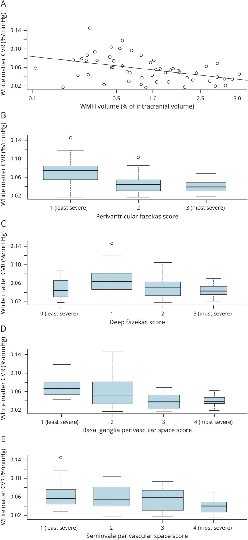
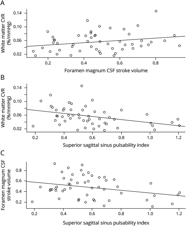
Results: Fifty-three of 60 patients completed the study with a full data set (age 68.0% ± 8.8 years, 74% male, 75% hypertensive). After controlling for age, sex, and systolic blood pressure, lower white matter CVR was associated with higher WMH volume (-0.01%/mm Hg per log10 increase in WMH volume, p = 0.02), basal ganglia PVS (-0.01%/mm Hg per point increase in the PVS score, p = 0.02), and higher venous pulsatility (superior sagittal sinus -0.03%/mm Hg, p = 0.02, per unit increase in the pulsatility index) but not with CBF (p = 0.58). Lower foramen magnum CSF stroke volume was associated with worse white matter CVR (0.04%/mm Hg per mL increase in stroke volume, p = 0.04) and more severe basal ganglia PVS (p = 0.09).
Conclusions: Lower CVR, higher venous pulsatility, and lower foramen magnum CSF stroke volume indicate that dynamic vascular dysfunctions underpin PVS dysfunction and WMH development. Further exploration of microvascular dysfunction and CSF dynamics may uncover new mechanisms and intervention targets to reduce SVD lesion development, cognitive decline, and stroke.
Copyright © 2020 The Author(s). Published by Wolters Kluwer Health, Inc. on behalf of the American Academy of Neurology.
Figures
Comment in
-
Cerebral small vessel disease: Moving closer to hemodynamic function.Neurology. 2020 May 26;94(21):909-910. doi: 10.1212/WNL.0000000000009477. Epub 2020 May 4. Neurology. 2020. PMID: 32366538 No abstract available.
References
Publication types
MeSH terms
Grants and funding
LinkOut - more resources
Full Text Sources
Other Literature Sources
Research Materials