Review
. 2020 Jun 22;88(7):e00930-19.
doi: 10.1128/IAI.00930-19. Print 2020 Jun 22.

Biting Off What Can Be Chewed: Trogocytosis in Health, Infection, and Disease

Affiliations
Review

Biting Off What Can Be Chewed: Trogocytosis in Health, Infection, and Disease

Akhila Bettadapur et al. Infect Immun. .

Abstract

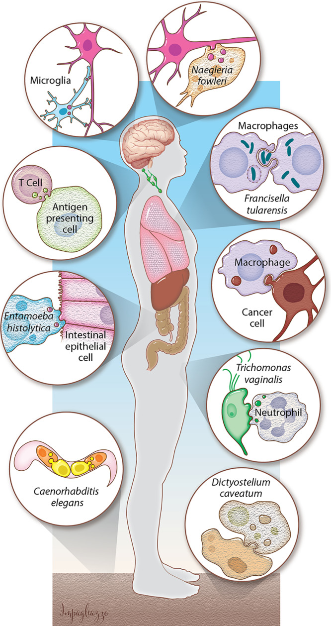
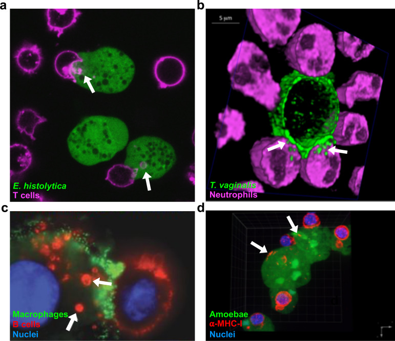
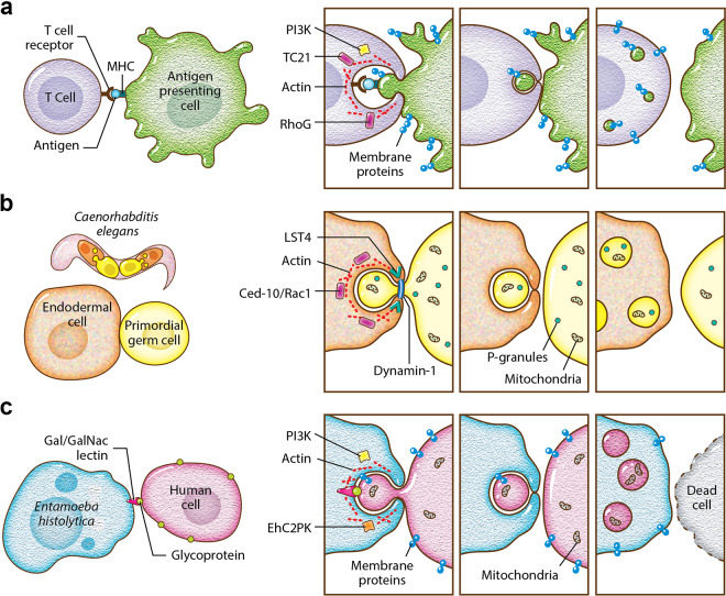
Trogocytosis is part of an emerging, exciting theme of cell-cell interactions both within and between species, and it is relevant to host-pathogen interactions in many different contexts. Trogocytosis is a process in which one cell physically extracts and ingests "bites" of cellular material from another cell. It was first described in eukaryotic microbes, where it was uncovered as a mechanism by which amoebae kill cells. Trogocytosis is potentially a fundamental form of eukaryotic cell-cell interaction, since it also occurs in multicellular organisms, where it has functions in the immune system, in the central nervous system, and during development. There are numerous scenarios in which trogocytosis occurs and an ever-evolving list of functions associated with this process. Many aspects of trogocytosis are relevant to microbial pathogenesis. It was recently discovered that immune cells perform trogocytosis to kill Trichomonas vaginalis parasites. Additionally, through trogocytosis, Entamoeba histolytica acquires and displays human cell membrane proteins, enabling immune evasion. Intracellular bacteria seem to exploit host cell trogocytosis, since they can use it to spread from cell to cell. Thus, a picture is emerging in which trogocytosis plays critical roles in normal physiology, infection, and disease.

Keywords: Entamoeba; Francisella; Trichomonas; cell death; complement; macrophages; neutrophils; phagocytosis; trogocytosis.


Figures

FIG 1
FIG 1
Trogocytosis is a broad, developing concept. In the central nervous system, microglia use trogocytosis to remodel neuronal synapses, and the parasite N. fowleri kills human cells through trogocytosis. Immune cells take bites out of other human cells. Bacteria such as F. tularensis exploit trogocytosis/merocytophagy to spread between cells. Macrophages can perform trogocytosis to kill antibody-opsonized cells. E. histolytica kills human cells by performing trogocytosis. Neutrophils kill T. vaginalis through trogocytosis. Primordial germ cells in C. elegans are nibbled by endodermal cells. D. caveatum kills other Dictyostelium species through trogocytosis. (Courtesy of Anita Impagliazzo, reproduced with permission.)
FIG 2
FIG 2
Examples of trogocytosis within and between species. (a) E. histolytica kills human cells through trogocytosis. E. histolytica is stained with cell tracker green, and human Jurkat T cell membranes are stained with DiD (pink). Arrows, ingested bites. (b) Neutrophils kill T. vaginalis through trogocytosis. T. vaginalis membranes are stained with streptavidin-488 (green), and neutrophils are stained with cell tracker deep red (pink). Arrows, ingested bites. (c) Macrophages can perform trogocytosis to kill antibody-opsonized cells. Macrophages are stained with anti-CD45 (green), Raji B cells are opsonized with trastuzumab (red), and nuclei are stained with Hoechst stain (blue). Arrows, ingested bites. (d) E. histolytica acquires and displays human cell membrane proteins through trogocytosis. E. histolytica is stained with cell tracker green, human anti-MHC-I is shown in red, and nuclei are stained with 4′,6′-diamidino-2-phenylindole (DAPI) (blue). Arrows, acquired MHC-I. (Reprinted from references , , and with permission.)
FIG 3
FIG 3
The molecular mechanism underlying trogocytosis. (a) T cell trogocytosis. T cell receptors engage with antigen bound by MHC. The small GTPases TC21 and RhoG play roles in trogocytosis, along with PI3K and actin. Membrane proteins from the antigen-presenting cell are ultimately displayed on the T cell. The cells separate and remain viable. (b) C. elegans endodermal cell trogocytosis. The small GTPase CED-10/Rac1 plays a role in trogocytosis, along with actin, Lst-4/SNX9, and dynamin-1. Lst-4/SNX9 has a role in membrane bending, and dynamin-1 has a role in membrane scission. Some P-granules and mitochondria are removed from the primordial germ cell. The cells separate without cell death, and after trogocytosis, the primordial germ cell is smaller and contains fewer P-granules and mitochondria. (c) E. histolytica trogocytosis. Glycoproteins on the human cell surface are engaged by the Gal/GalNAc lectin. The kinase EhC2PK plays a role in trogocytosis, together with PI3K and actin. Membrane proteins from the human cell are ultimately displayed on the amoeba. The cells separate once the human cell is killed. (Courtesy of Anita Impagliazzo, reproduced with permission.)

References

    1. Dance A. 2019. Core concept: cells nibble one another via the under-appreciated process of trogocytosis. Proc Natl Acad Sci U S A 116:17608–17610. doi:10.1073/pnas.1912252116. - DOI - PMC - PubMed
    1. Ralston KS. 2015. Taking a bite: amoebic trogocytosis in Entamoeba histolytica and beyond. Curr Opin Microbiol 28:26–35. doi:10.1016/j.mib.2015.07.009. - DOI - PMC - PubMed
    1. Batista FD, Iber D, Neuberger MS. 2001. B cells acquire antigen from target cells after synapse formation. Nature 411:489–494. doi:10.1038/35078099. - DOI - PubMed
    1. Miyake K, Shiozawa N, Nagao T, Yoshikawa S, Yamanishi Y, Karasuyama H. 2017. Trogocytosis of peptide–MHC class II complexes from dendritic cells confers antigen-presenting ability on basophils. Proc Natl Acad Sci U S A 114:1111–1116. doi:10.1073/pnas.1615973114. - DOI - PMC - PubMed
    1. Li K-J, Wu C-H, Shen C-Y, Kuo Y-M, Yu C-L, Hsieh S-C. 2016. Membrane transfer from mononuclear cells to polymorphonuclear neutrophils transduces cell survival and activation signals in the recipient cells via anti-extrinsic apoptotic and MAP kinase signaling pathways. PLoS One 11:e0156262. doi:10.1371/journal.pone.0156262. - DOI - PMC - PubMed

Publication types

Substances