. 2020 May 4;10(1):7443.
doi: 10.1038/s41598-020-63619-y.

Snakes elicit specific neural responses in the human infant brain

Affiliations

Snakes elicit specific neural responses in the human infant brain

J Bertels et al. Sci Rep. .

Abstract

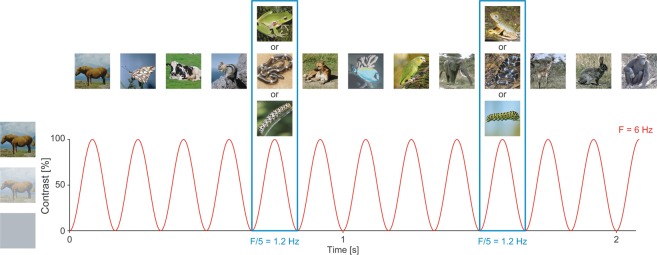
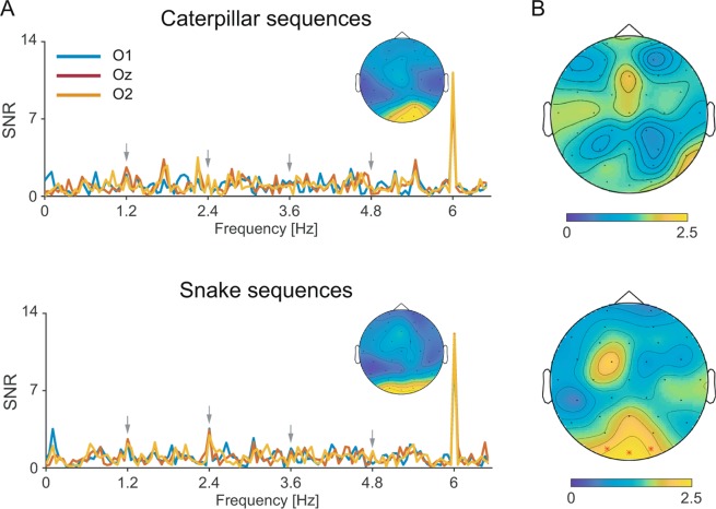
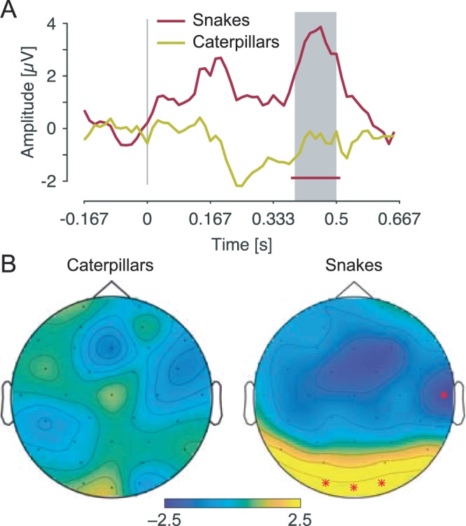
Detecting predators is essential for survival. Given that snakes are the first of primates' major predators, natural selection may have fostered efficient snake detection mechanisms to allow for optimal defensive behavior. Here, we provide electrophysiological evidence for a brain-anchored evolved predisposition to rapidly detect snakes in humans, which does not depend on previous exposure or knowledge about snakes. To do so, we recorded scalp electrical brain activity in 7- to 10-month-old infants watching sequences of flickering animal pictures. All animals were presented in their natural background. We showed that glancing at snakes generates specific neural responses in the infant brain, that are higher in amplitude than those generated by frogs or caterpillars, especially in the occipital region of the brain. The temporal dynamics of these neural responses support that infants devote increased attention to snakes than to non-snake stimuli. These results therefore demonstrate that a single fixation at snakes is sufficient to generate a prompt and large selective response in the infant brain. They argue for the existence in humans of an inborn, brain-anchored mechanism to swiftly detect snakes based on their characteristic visual features.


Conflict of interest statement

The authors declare no competing interests.

Figures

Figure 1
Figure 1
Schematic illustration of the experimental paradigm used. Animal pictures were presented by sinusoidal contrast modulation at a rate of 6 per second (F = 6 Hz). Snake, frog or caterpillar pictures were presented every fifth stimulus (F = 6/5 = 1.2 Hz), in different trial sequences. Snake and frog sequences were used in the main study; snake and caterpillar sequences were used in the control study. The pictures differed in terms of color, viewpoint and lighting conditions. Snake, frog, caterpillar and other animal pictures were equalized in terms of luminance and contrast across the whole set. For copyright reasons, the pictures of snakes, frogs and caterpillars displayed are different than those used in the actual experiment (originally coming from Vanessa LoBue’s personal database), but the degree of variability across images is respected. Most of other animal pictures come from CalPhotos (https://calphotos.berkeley.edu/fauna).
Figure 2
Figure 2
Frequency-domain representation of frog and snake-selective responses during fast periodic visual stimulation (left and right panels, respectively). (A) SNR spectra of each occipital electrode (O1, Oz, O2) and topographical maps of SNR at the base frequency (6 Hz). Asterisks indicate significant oddball responses. (B) Topographical maps of SNR at each harmonic of the oddball frequency (1.2, 2.4, 3.6, and 4.8 Hz; left part), and of SNR averaged on these first four harmonics (right part). Asterisks indicate significant responses at specific channels.
Figure 3
Figure 3
Time-domain representation of frog and snake-selective responses during fast periodic visual stimulation. (A) Grand averages of the notch-filtered EEG responses relative to the onset of the frog and snake stimuli, at O2. The red line below the waveforms represents the time-points at which the signal significantly deviates from baseline after snake pictures. The grey area indicates the time-window at which the signal significantly differs between frog and snake pictures. Note that the amplitude scale is only approximate due to the epoch-normalization scheme used (see methods). (B) Topographical maps of the P400 evoked by frog and snake stimuli in the time-window at which the signal significantly differs between them (i.e., 420–490 post-stimulus onset).
Figure 4
Figure 4
Frequency-domain representation of caterpillar and snake-selective responses during fast periodic visual stimulation (left and right panels, respectively). (A) SNR spectra of each occipital electrode (O1, Oz, O2) and topographical maps of SNR at the base frequency (6 Hz). (B) Topographical maps of SNR averaged on the first four harmonics.
Figure 5
Figure 5
Time-domain representation of caterpillar and snake-selective responses during fast periodic visual stimulation. (A) Grand averages of the notch-filtered EEG responses relative to the onset of the caterpillar and snake stimuli, at O2. The red line below the waveforms represents the time-points at which the signal significantly deviates from baseline after snake pictures. The grey area indicates the time-window at which the signal significantly differs between caterpillar and snake pictures. Note that the amplitude scale is only approximate due to the epoch-normalization scheme used (see methods). (B) Topographical maps of the P400 evoked by caterpillar and snake stimuli in the time-window at which the signal significantly differs between both (i.e., 390–500 ms post-stimulus onset).

References

    1. Isbell LA. Snakes as agents of evolutionary change in primate brains. J. Hum. Evol. 2006;51:1–35. doi: 10.1016/j.jhevol.2005.12.012. - DOI - PubMed
    1. Isbell, L. A. The Fruit, the Tree, and the Serpent. (Harvard University Press, 2009).
    1. Öhman A, Mineka S. The malicious serpent: Snakes as a prototypical stimulus for an evolved module of fear. Curr. Dir. Psychol. Sci. 2003;12:5–9. doi: 10.1111/1467-8721.01211. - DOI
    1. Van Le Q, et al. Pulvinar neurons reveal neurobiological evidence of past selection for rapid detection of snakes. Proc. Natl. Acad. Sci. 2013;110:19000–19005. doi: 10.1073/pnas.1312531110. - DOI - PMC - PubMed
    1. Soares SC, Lindström B, Esteves F, Öhman A. The hidden snake in the grass: Superior detection of snakes in challenging attentional conditions. Plos One. 2014;9:e114724. doi: 10.1371/journal.pone.0114724. - DOI - PMC - PubMed

Publication types