. 2020 May 4;11(1):2187.
doi: 10.1038/s41467-020-16121-y.

A microsporidian impairs Plasmodium falciparum transmission in Anopheles arabiensis mosquitoes

Affiliations

A microsporidian impairs Plasmodium falciparum transmission in Anopheles arabiensis mosquitoes

Jeremy K Herren et al. Nat Commun. .

Abstract

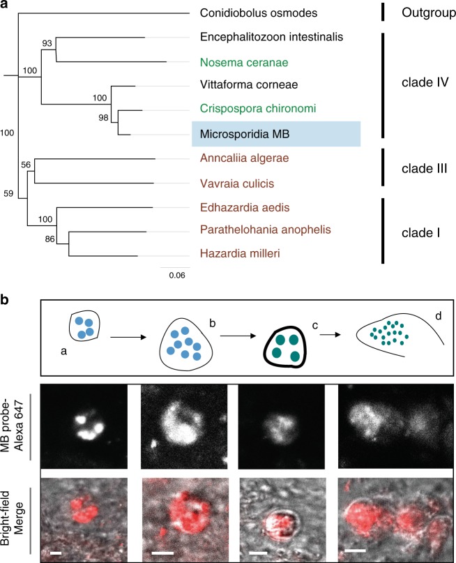
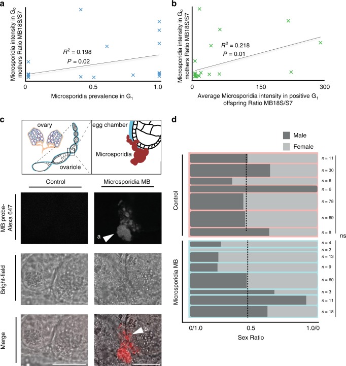
A possible malaria control approach involves the dissemination in mosquitoes of inherited symbiotic microbes to block Plasmodium transmission. However, in the Anopheles gambiae complex, the primary African vectors of malaria, there are limited reports of inherited symbionts that impair transmission. We show that a vertically transmitted microsporidian symbiont (Microsporidia MB) in the An. gambiae complex can impair Plasmodium transmission. Microsporidia MB is present at moderate prevalence in geographically dispersed populations of An. arabiensis in Kenya, localized to the mosquito midgut and ovaries, and is not associated with significant reductions in adult host fecundity or survival. Field-collected Microsporidia MB infected An. arabiensis tested negative for P. falciparum gametocytes and, on experimental infection with P. falciparum, sporozoites aren't detected in Microsporidia MB infected mosquitoes. As a microbe that impairs Plasmodium transmission that is non-virulent and vertically transmitted, Microsporidia MB could be investigated as a strategy to limit malaria transmission.


Conflict of interest statement

The authors declare no competing interests.

Figures

Fig. 1
Fig. 1. A microsporidian associated with An. arabiensis populations in Kenya.
a 18S rDNA-based phylogeny reveals that Microsporidia MB are in clade IV of the Microsporidia. Labeled in brown are the microsporidian species known from mosquitoes. In green are microsporidians associated with other insect groups. Microsporidia MB 18S rDNA gene sequence deposited in GenBank (accession number MT160806[https://www.ncbi.nlm.nih.gov/nuccore/MT160806]) and in Source Data file. b Fluorescence in situ hybridization (FISH) staining of the diplokaryotic stages of Microsporidia MB merogony (a, b) and spore capsule formation (c, d) in An. arabiensis larval gut epithelial tissues. Red in the merge images is the MB probe-Alexa 647 FISH probe targeting Microsporidia MB. The scale bar is 1.5 μm. Representative images as observed in four independent experiments.
Fig. 2
Fig. 2. Seasonal variation in Microsporidia MB prevalence in two sampling sites.
Weekly precipitation values given are based on earth observation and are shown in dark blue. Light blue bars indicate the prevalence of Microsporidia MB, monitored in weekly collections. Overall, 136 out of 2274 (6%) female An. arabiensis were found to be infected with Microsporidia MB in Mwea and 159 out of 1996 (8%) female An. arabiensis in Ahero. Source data are provided as a Source Data file.
Fig. 3
Fig. 3. Microsporidia MB is maternally transmitted and does not bias sex ratio.
a The vertical transmission efficiency of Microsporidia MB to G1 depends on the G0 (maternal) Microsporidia MB intensity. In a linear regression, the slope is significantly non-zero (P = 0.02, F = 6.169, and n = 27). b The intensity of Microsporidia MB in G1 offspring is correlated to G0 maternal intensity, with a linear regression the slope that is significantly non-zero (P = 0.01, F = 6.98 and n = 27). c Fluorescence microscopy with a Microsporidia MB specific FISH probe (MB probe) indicates that Microsporidia MB (a) is localized to the posterior of developing vitellogenic egg chambers (b) in An. arabiensis. Scale bar, 20 μm. Representative images as observed in three independent experiments. d The sex ratio of broods from Microsporidia MB infected mothers does not differ significantly from Microsporidia MB uninfected mothers suggesting there is no sex ratio distortion (Unpaired two-tailed t-test, P = 0.32, t = 1.02, df=13). Dashed line reflects overall sex ratio (Uninfected control = 100:108 or 52% female, Microsporidia MB = 56:64 or 54% female). Column widths reflect sample sizes Source data are provided as a Source Data file.
Fig. 4
Fig. 4. Microsporidia MB impairs parasite development in An. arabiensis after direct membrane feeding assay challenge with P. falciparum.
The Plasmodium infection rate in Microsporidia MB positive and Microsporidia MB negative mosquitoes was determined by qPCR. a The head and thorax Plasmodium infection rate, reflecting presence of sporozoites, in Microsporidia MB positive and Microsporidia MB negative mosquitoes. There was a significant absence of co-infected mosquitoes (two-tailed fisher exact test, P = 0.02 n = 178). b Plasmodium intensity in An. arabiensis heads and thoraxes, quantified by qPCR. c The abdomen Plasmodium infection rate, reflecting presence of oocysts, in Microsporidia MB positive and Microsporidia MB negative mosquitoes. There was a significant absence of co-infected mosquitoes (two-tailed fisher exact test, P = 0.04, n = 140). d Plasmodium intensity in An. arabiensis abdomens, quantified by qPCR. Data shown in (a, b, c, d) are pooled from replicate experiments carried out using different gametocyte donors (for each donor the numbers of Plasmodium positive An. arabiensis is shown in red and Microsporidia MB positives An. arabiensis is shown in blue). Each data point is an individual An. arabiensis mosquito. Source data are provided as a Source Data file.
Fig. 5
Fig. 5. Microsporidia MB does not overtly decrease host fitness.
a Microsporidia MB harboring wild-caught An. arabiensis did not lay significantly less eggs than uninfected counterparts (two-tailed Mann–Whitney test, P = 0.18). The black line indicates the mean and error bars reflect SEM. b The G1 larval progeny of Microsporidia MB infected wild-caught females develop significantly faster than uninfected counterparts. c The larval development of Microsporidia MB infected An. arabiensis is on average 1.1, 1.4, and 1.3 days less than uninfected controls in three independent experiments (Two-tailed Mann–Whitney test, P < 0.0001, P = 0.03, P < 0.0001, error bars reflect SEM). Mean values are shown above the scatter dot plots. Data shown in (b) and (c) are from the same three independent experiments. d The survival of adult G1 progeny of Microsporidia MB infected wild-caught females is not significantly different from uninfected counterparts, one experiment (ii) shown of three independent experiments (i-iii); (i, P = 0.86, n > 9, ii, P = 0.97, n > 41, ii, P = 0.06 n > 50, n denotes the minimum number of mosquitoes per condition). e The intensity of Microsporidia MB across different developmental stages. The intensity of Microsporidia MB is highly variable in larvae, lower in young adults but increases as adult An. arabiensis females age. Boxplot boundaries reflect the inter-quartile range. The horizontal bar is the median and whiskers extend to 1.5 times the inter-quartile range beyond the boxplot. Each datapoint represents an individual mosquito from a total of five (L4-day 2), four (day 8) and three (day 10 and 15) independent experiments. Source data are provided as a Source Data file.

References

    1. WHO. World Malaria Report (World Health Organization, 2018).
    1. Moreira LA, et al. A Wolbachia symbiont in Aedes aegypti limits infection with dengue, Chikungunya, and Plasmodium. Cell. 2009;139:1268–1278. doi: 10.1016/j.cell.2009.11.042. - DOI - PubMed
    1. Bian G, Xu Y, Lu P, Xie Y, Xi Z. The endosymbiotic bacterium Wolbachia induces resistance to dengue virus in Aedes aegypti. PLoS Pathog. 2010;6:e1000833. doi: 10.1371/journal.ppat.1000833. - DOI - PMC - PubMed
    1. Walker T, et al. The wMel Wolbachia strain blocks dengue and invades caged Aedes aegypti populations. Nature. 2011;476:450–453. doi: 10.1038/nature10355. - DOI - PubMed
    1. Hoffmann AA, et al. Successful establishment of Wolbachia in Aedes populations to suppress dengue transmission. Nature. 2011;476:454–457. doi: 10.1038/nature10356. - DOI - PubMed

Publication types