

Review
doi: 10.1038/s41433-020-0902-4.
Epub 2020 May 4.
Congenital focal abnormalities of the retina and retinal pigment epithelium
Affiliations
- PMID: 32367006
- PMCID: PMC7784997
- DOI: 10.1038/s41433-020-0902-4
Item in Clipboard
Review
Congenital focal abnormalities of the retina and retinal pigment epithelium
Eye (Lond).
2020 Nov.
Abstract
This paper reviews the published literature on a group of developmental disorders of the retina and retinal pigment epithelium which result in focal abnormalities in one or both eyes. They are often asymptomatic, found on routine examination and are generally non-progressive. Some are associated with other systemic abnormalities.
Conflict of interest statement
The authors declare that they have no conflict of interest.
Figures

a A solitary flat, pigmented, round CHRPE lesion with a ring of hypopigmentation within the margins. b An example of lacunae, a hypopigmented, punched-out patch within another CHRPE lesion.

a The lesions are smaller centrally and enlarge in size toward the periphery, resembling animal footprints. b The lesions tend to occur in one sector of the retina, with apex of the sector toward the optic nerve, and tend to be black in the centre and become lighter in colour towards the periphery.

a Widespread patchy chalky white lesions of the left eye in one patient. b Fundus autofluorescence in the same eye shows that albinotic spots appear hypoautofluorescent. c Diffuse albinotic spots of the left eye in a different patient. d In the same patient, albinotic spots demonstrates hyperautofluorescence on fundus autofluorescence.

a, b RPE lesions with irregular border and variable pigmentation in two different patients with FAP.
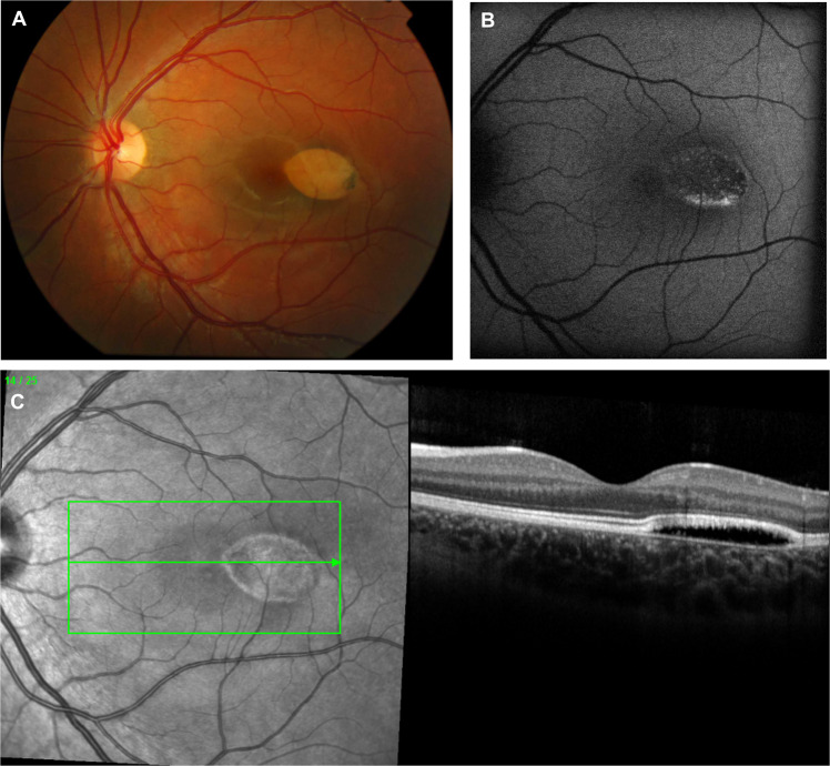
a A typical lesion is present in the temporal macula of the left eye straddling the horizontal raphe, with a tail pointing towards the fovea. b Fundus autofluorescence of the same lesion demonstrates a mostly hypoautofluorescent lesion with a rim of hyperautofluorescence along the inferior margin. c Optical coherence tomography through the lesion demonstrates a subretinal cleft with elevation of the neurosensory retina.

a A focal, well-circumscribed, jet-black lesion just temporal to the fovea in the left eye. b Fundus autofluorescence of the same lesion demonstrates hypoautofluorescence from heavy pigmentation in the lesion. c Optical coherence tomography shows prominent hyperreflectivity in the area of the lesion with abrupt and complete posterior optical shadowing.

a, b Two eyes of the same infant with a family history of neurofibromatosis 2 (NF2) showing evidence of bilateral combined hamartomas, indicating that the infant also has NF2. c An ill-defined, lightly pigmented, elevated mass located in the macula associated with epiretinal membrane and vascular tortuosity. d Late phase fluorescein angiogram of the same lesion demonstrates diffuse late punctate leakage within the lesion. e Optical coherence tomography through the lesion shows a diffusely thickened retina with intraretinal fluid and disorganisation of the inner and outer retinal architecture.

a, b are fundus photos of a patient with TSC with bilateral lesions. c A solitary elevated mass with internal calcification in the fundus of the right eye in a patient without a diagnosis of TSC. d Fundus autofluorescence of the same lesion demonstrates hyperautofluorescence in areas of internal calcification. e Optical coherence tomography scanning through the lesion demonstrates an abrupt transition from normal retina to an elevated mass with disorganisation of the inner retina and internal moth-eaten empty spaces that correlate with intratumoral calcification, in addition to complete posterior shadowing.

a Normal-sized right eye of a patient with Aicardi syndrome. b Microphthalmic left eye in the same patient. c Left eye demonstrates persistent pupillary membrane. d Right eye demonstrates optic nerve coloboma and multiple well-circumscribed, round chorioretinal lesions in the posterior pole, consistent with chorioretinal lacunae. One large lacunae beneath the optic disc appears to have subretinal fluid, confirmed on OCT. e Optical coherence tomography reveals an outer retinal cavity filled with subretinal fluid over one of the lacunae. f Left eye shows a complete funnel retinal detachment.

a Left eye of a patient with KIF11 disease demonstrates chorioretinopathy with atrophy, local vessel attenuation and pigment clumping inferior to the macula. b Fundus autofluorescence of the same eye demonstrates hypoautofluorescence in areas of chorioretinopathy consistent with RPE atrophy. c Optical coherence tomography shows loss of retinal lamination and absence of outer retinal architecture within areas of the lesion.
References
-
- Gass JD. Focal congenital anomalies of the retinal pigment epithelium. Eye. 1989;3(Pt 1):1–18. - PubMed
-
- Buettner H. Congenital hypertrophy of the retinal pigment epithelium. Am J Ophthalmol. 1975;79:177–89. - PubMed
-
- Coleman P, Barnard NAS. Congenital hypertrophy of the retinal pigment epithelium: prevalence and ocular features in the optometric population. Ophthalmic Physiol Opt. 2007;27:547–55. - PubMed
-
- Shields CL, Mashayekhi A, Ho T, Cater J, Shields JA. Solitary congenital hypertrophy of the retinal pigment epithelium: clinical features and frequency of enlargement in 330 patients. Ophthalmology. 2003;110:1968–76. - PubMed
-
- Champion R, Daicker BC. Congenital hypertrophy of the pigment epithelium: light microscopic and ultrastructural findings in young children. Retina. 1989;9:44–8. - PubMed
