

Review
doi: 10.1038/s41590-020-0677-6.
Epub 2020 May 4.
Autoimmunity and organ damage in systemic lupus erythematosus
Affiliations
- PMID: 32367037
- PMCID: PMC8135909
- DOI: 10.1038/s41590-020-0677-6
Item in Clipboard
Review
Autoimmunity and organ damage in systemic lupus erythematosus
Nat Immunol.
2020 Jun.
Abstract
Impressive progress has been made over the last several years toward understanding how almost every aspect of the immune system contributes to the expression of systemic autoimmunity. In parallel, studies have shed light on the mechanisms that contribute to organ inflammation and damage. New approaches that address the complicated interaction between genetic variants, epigenetic processes, sex and the environment promise to enlighten the multitude of pathways that lead to what is clinically defined as systemic lupus erythematosus. It is expected that each patient owns a unique 'interactome', which will dictate specific treatment.
Conflict of interest statement
Competing interests
The author declares no competing interests.
Figures
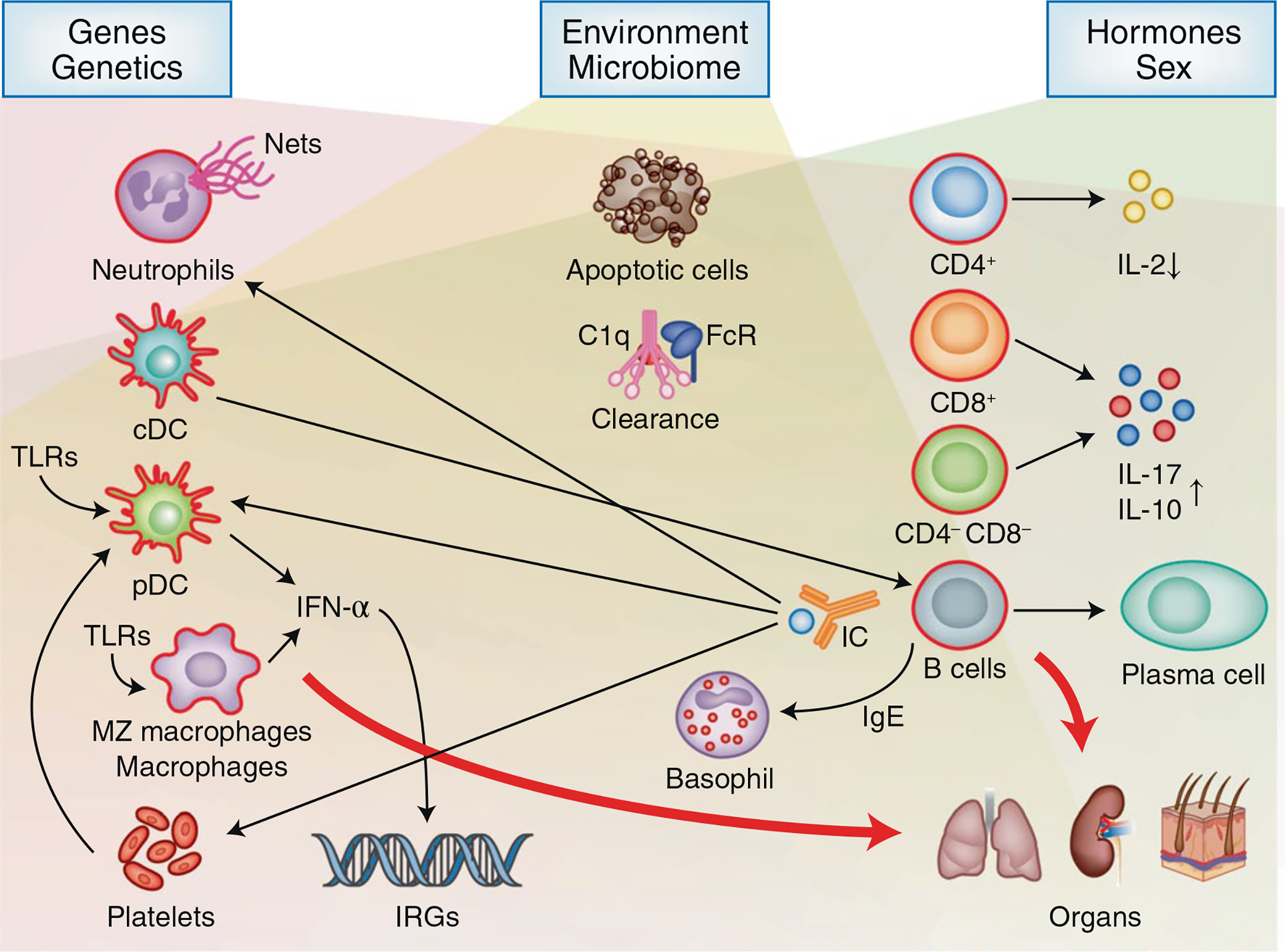
Genetic, environmental and hormonal factors act on various elements of the innate and adaptive immune responses. Gene copy variants (for example, C4 and FCR) and SNPs influence the expression of many genes involved in the immune response as well as in the control of organ damage (for example, APOL1 and KLK). Environmental factors, including UV light, drugs and products of the microbiome, alter T and B cell responses and the functions of innate cells by stimulating TLRs. Hormones and genes defined by the X chromosome contribute to disease expression by altering the function of lymphocytes and of cells of the innate immune response. The involved factors lead eventually to loss of tolerance of B and T cells to autoantigens, which are present in abundance because of both increased rates of apoptosis and defects in mechanisms responsible for their clearance. The T cell response to antigen is aberrant in and late signaling events (Fig. 2) and results in misbalanced levels of cytokines, including decreased IL-2 and increased IL-17 production. T cells, through distinct pathways, also acquire a greater ability to invade tissues and contribute to the inflammatory response. B cells, in response to cognate and non-cognate (cytokines) interactions with T cells, produce antibodies. Antibodies enter tissues directly or in the form of immune complexes (IC), which contribute to tissue inflammation. Cells of the innate immune response, under the influence of the involved pathogenic factors, produce cytokines (including IFN-α) or, through the direct interaction with lymphocytes, contribute significantly to the inflammatory organ-damaging response. The clinical heterogeneity of the disease is highly correlated with the multitude of the pathways that lead to organ injury. Although several pathways operate in each individual, the relative contribution of each pathway varies from person to person. Finally, local factors dictate which organ will be afflicted by the autoinflammatory response.
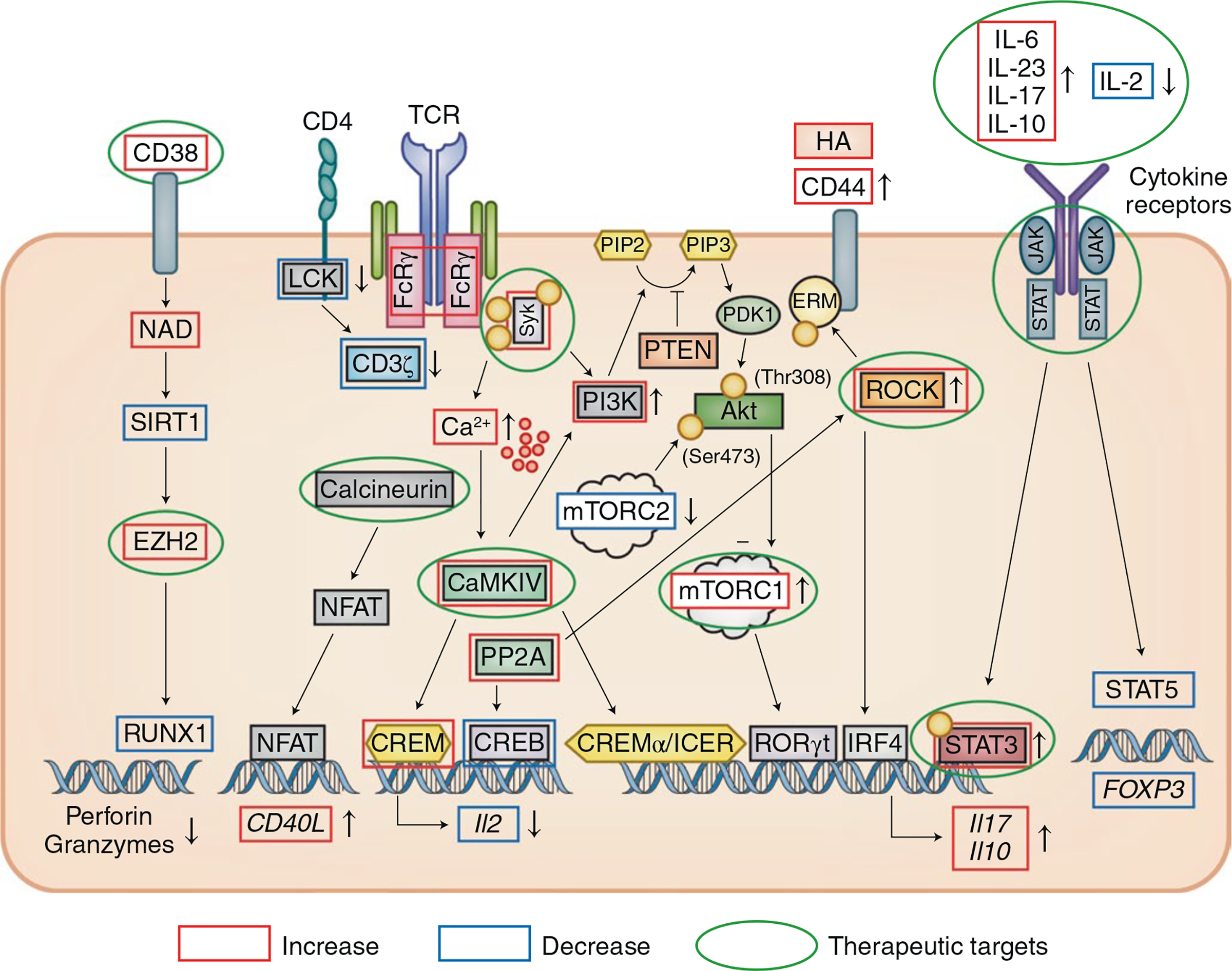
Engagement of the CD3 complex either by autoantigen or circulating CD3/TCR antibodies results in an increased intracytoplasmic calcium response. The CD3 complex is rewired, with the CD3ζ chain replaced with the FcRγ chain, which recruits the kinase Syk. The calcium-requiring kinase CaMKIV enhances the binding of CREMα/ICER to the Il2 and Il17 or Il10 promoters to suppress and enhance their expression, respectively. Calcineurin also dephosphorylates the transcription factor NFAT, which binds to the promoter of CD40L and increases its expression. Phosphatase PP2A is increased in T cells with diverse effects: in Treg cells, it dephosphorylates mTORC1 and promotes Treg cell function and, in effector T cells, it enhances the binding of IRF4 to the Il17 promoter and it dephosphorylates p-CREB. PP2A also promotes ROCK activity, which phosphorylates ERM, which in turn enhances the ability of CD44 to bind its ligand hyaluronic acid (HA, expressed in the kidney and other tissues). IL-2 signaling is defective with decreased amounts of p-STAT5, whereas IL-6 signaling is increased with increased binding of p-STAT3 to the Il17 promoter. An increased proportion of CD8+ T cells express the ectonucleotidase CD38, which suppresses the level of NAD and the activity of the deacetylase SIRT1, which in turn enhances the activity of the histone methyltransferase EZH2. As indicated by the green circles, a number of the molecules have been considered as therapeutic targets: Syk, ROCK, calcineurin, EZH2, IL-17, IL-23, JAK and mTOR as well as IL-2, which can be replenished with low doses.
References
-
- Durcan L, O’Dwyer T & Petri M Management strategies and future directions for systemic lupus erythematosus in adults. Lancet 393, 2332–2343 (2019). - PubMed
-
- Dörner T & Furie R Novel paradigms in systemic lupus erythematosus. Lancet 393, 2344–2358 (2019). - PubMed
-
- Tsokos GC Systemic lupus erythematosus. N. Engl. J. Med. 365, 2110–2121 (2011). - PubMed
-
- Deng Y & Tsao BP Updates in lupus genetics. Curr. Rheumatol. Rep. 19, 68 (2017). - PubMed
Publication types
MeSH terms
Grants and funding
LinkOut - more resources
Full Text Sources
Other Literature Sources
Medical
