Review
. 2020 Apr 23:15:885-899.
doi: 10.2147/COPD.S236640. eCollection 2020.

Chitinases and Chitinase-Like Proteins in Obstructive Lung Diseases - Current Concepts and Potential Applications

Affiliations
Review

Chitinases and Chitinase-Like Proteins in Obstructive Lung Diseases - Current Concepts and Potential Applications

Natalia Przysucha et al. Int J Chron Obstruct Pulmon Dis. .

Abstract

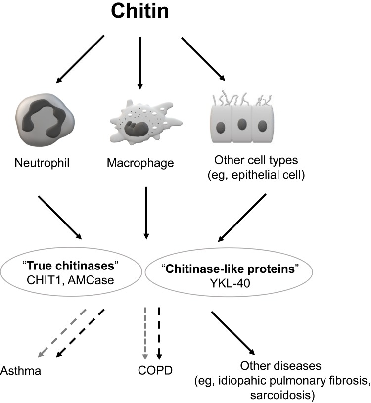
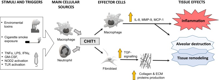
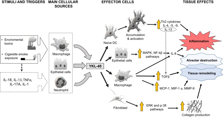
Chitinases, enzymes that cleave chitin's chain to low molecular weight chitooligomers, are widely distributed in nature. Mammalian chitinases belong to the 18-glycosyl-hydrolase family and can be divided into two groups: true chitinases with enzymatic activity (AMCase and chitotriosidase) and chitinase-like proteins (CLPs) molecules which can bind to chitin or chitooligosaccharides but lack enzymatic activity (eg, YKL-40). Chitinases are thought to be part of an innate immunity against chitin-containing parasites and fungal infections. Both groups of these hydrolases are lately evaluated also as chemical mediators or biomarkers involved in airway inflammation and fibrosis. The aim of this article is to present the current knowledge on the potential role of human chitinases and CLPs in the pathogenesis, diagnosis, and course of obstructive lung diseases. We also assessed the potential role of chitinase and CLPs inhibitors as therapeutic targets in chronic obstructive pulmonary disease and asthma.

Keywords: AMCase; CHIT1; COPD; YKL-40; asthma; chitotriosidase.


Conflict of interest statement

KG reports fees for lectures and travel expenses from Boehringer Ingelheim, Chiesi, AstraZeneca, Polpharma and Roche, outside the submitted work. RK reports personal fees and non-financial support from Boehringer Ingelheim, Chiesi, and AstraZeneca; and personal fees from Polpharma, outside the submitted work. The authors report no other conflicts of interest in this work.

Figures

Figure 1
Figure 1
Schematic depiction of chitin, chitinases and chitinase-like proteins (CLPs) involvement in lung diseases. Notes: Exposure to chitin may trigger the secretion of chitinases and CLPs from neutrophils, macrophages and other cells (solid-line black arrows show cell activation after chitin exposure and secretion of chitinases). Both groups of enzymes are thought to play a role in the pathogenesis of pulmonary diseases (eg, asthma and COPD). Black dashed arrows reflect a negative (ie, destructive) effect, while the grey dashed arrows show a possible positive (ie, protective) influence. See manuscript text for more details.
Figure 2
Figure 2
Schematic presentation of the hypothesized links between chitotriosidase (CHIT1) and the pathogenesis of lung inflammation and injury (eg, in COPD). Notes: The exposure to cigarette smoke and other factors may enhance CHIT1 expression and increase its production, mainly by lung macrophages and neutrophils. The impact of CHIT1 on lung cells is heterogeneous and include inflammation and tissue destruction, mediated by interleukin 8 (IL-8), metalloproteinase 9 (MMP-9) and monocyte chemoattractant protein 1 (MCP-1), as well as lung fibrosis induced by transforming growth factor-beta (TGF-β) pathway. However, the exact role of CHIT1 in the pathogenesis of lung diseases is still highly hypothetical. A large white arrow represents stimulation of macrophages and neutrophils by various factors, the thick grey arrows show the secretion of CHIT1 from the cellular sources, the thin black arrows depict the pathways of CHIT1 activity, the vertical yellow arrow represents the increase or enhancement.
Figure 3
Figure 3
Schematic representation of the supposed mechanisms involved in YKL-40-related emphysematous lung destruction, tissue inflammation and remodeling. Notes: The large white arrow represents stimulation of macrophages, epithelial cells and neutrophils by various factors, the thin dashed black arrow depicts an indirect effect of environmental factors, the thick grey arrows show the secretion of YKL-40 from the cellular sources, the thin black arrows depict the possible YKL-40 signaling pathways, the thin dashed grey arrow depicts a stimulatory effect of interleukin 13 (IL-13) on transforming growth factor-beta (TGFβ), the vertical yellow arrow represents the increase or enhancement. See manuscript text for more details.

Similar articles

Cited by

References

    1. Shahidi F, Abuzaytoun R. Chitin, chitosan, and co-products: chemistry, production, applications, and health effects. Adv Food Nutr Res. 2005;49:93–135. - PubMed
    1. Stern R. Go fly a chitin: the mystery of chitin and chitinases in vertebrate tissues. Front Biosci Landmark Ed. 2017;22:580–595. doi:10.2741/4504 - DOI - PubMed
    1. Sharp RG. A review of the applications of chitin and its derivatives in agriculture to modify plant-microbial interactions and improve crop yields. Agronomy. 2013;3(4):757–793. doi:10.3390/agronomy3040757 - DOI
    1. Kato Y, Onishi H, Machida Y. Application of chitin and chitosan derivatives in the pharmaceutical field. Curr Pharm Biotechnol. 2003;4(5):303–309. doi:10.2174/1389201033489748 - DOI - PubMed
    1. Yang TL. Chitin-based materials in tissue engineering: applications in soft tissue and epithelial organ. Int J Mol Sci. 2011;12(3):1936–1963. doi:10.3390/ijms12031936 - DOI - PMC - PubMed