. 2020 Apr 22:15:2733-2749.
doi: 10.2147/IJN.S249773. eCollection 2020.

Characterization and Cytotoxicity of Polyprenol Lipid and Vitamin E-TPGS Hybrid Nanoparticles for Betulinic Acid and Low-Substituted Hydroxyl Fullerenol in MHCC97H and L02 Cells

Affiliations

Characterization and Cytotoxicity of Polyprenol Lipid and Vitamin E-TPGS Hybrid Nanoparticles for Betulinic Acid and Low-Substituted Hydroxyl Fullerenol in MHCC97H and L02 Cells

Ran Tao et al. Int J Nanomedicine. .

Abstract

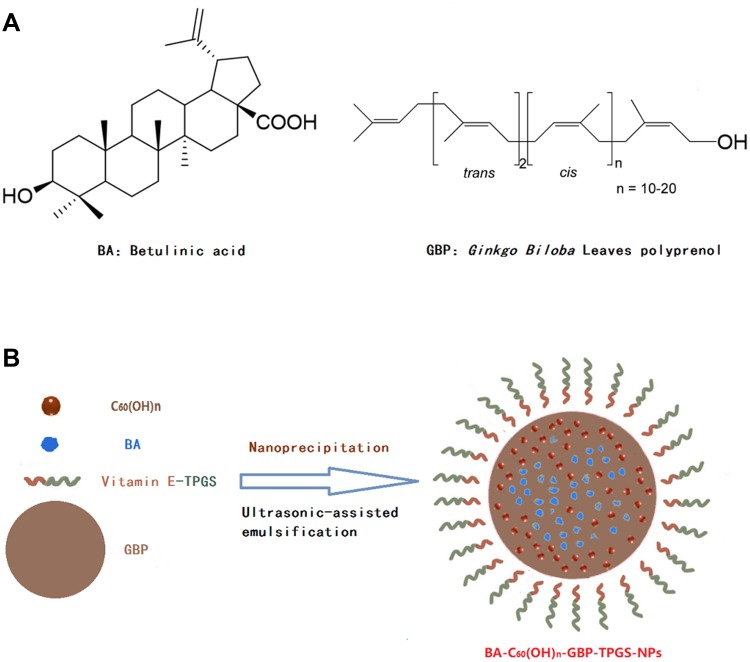
Background: This study demonstrated an innovative formulation including the polyprenol (GBP) lipid and vitamin E-TPGS hybrid nanoparticles (NPs) which was aimed to control the transfer of betulinic acid (BA) and low-substituted hydroxyl fullerenol (C60(OH)n). Additionally, it developed BA-C60(OH)n-GBP-TPGS-NPs delivery system and researched the anti-hepatocellular carcinoma (HCC) effects.

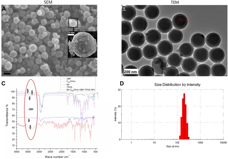
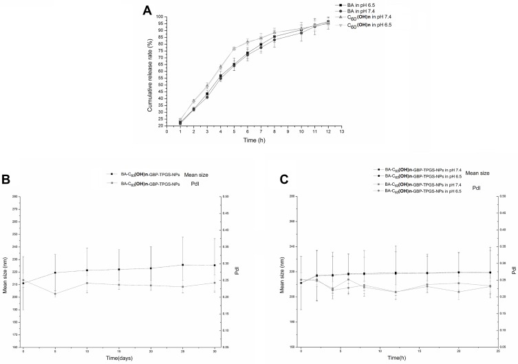
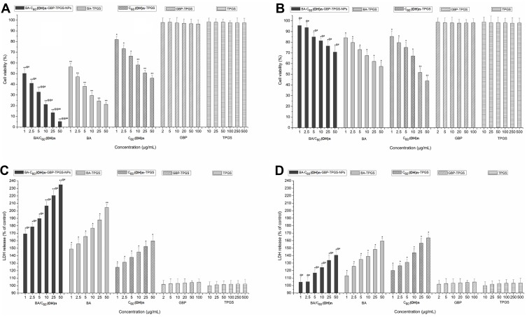
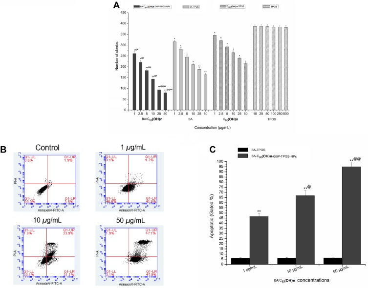
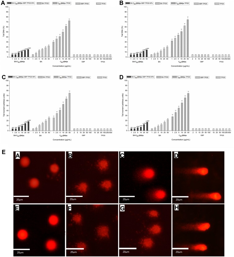
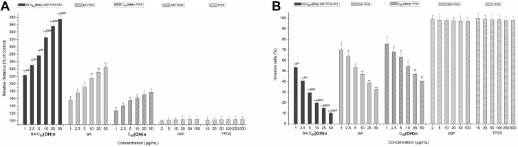
Materials and methods: The NPs were prepared by nanoprecipitation with ultrasonic-assisted emulsification (UAE) method. It was characterized by scanning electronic microscopy (SEM), transmission electron microscopy (TEM), FTIR spectrum, size distribution and zeta potential. Physical and chemical properties were evaluated through measurement of drug release, stability studies, drug loading efficiency (DE) and encapsulation efficiency (EE). Biological activities were evaluated through measurement of MTT assay, lactate dehydrogenase leakage assay (LDH), cell proliferation assays, cell apoptosis analysis, comet assay, wound healing assay, cell invasion and Western blot analysis.

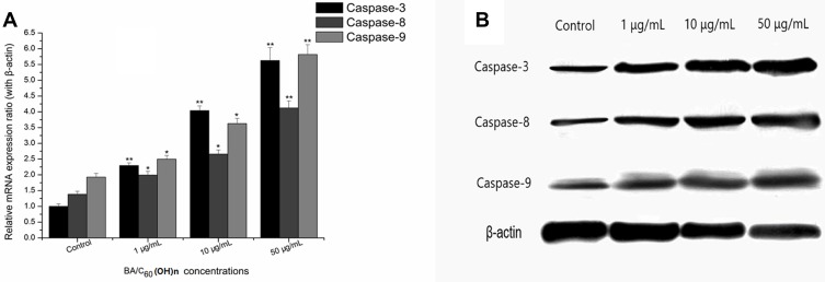
Results and conclusions: The NPs exhibited clear distribution characteristics, improved solubility and stability. BA and C60(OH)n for the NPs displayed a biphasic release pattern with sustained drug release properties. The mixture of C60(OH)n with different hydroxyl groups may have a certain effect on the stability of the NPs system itself. The NPs could effectively inhibit MHCC97H cell proliferation, migration and invasion in vitro. Combined use of C60(OH)n and BA in GBP lipids may improve the inhibit effect of C60(OH)n or BA against HCC cells and reduce cytotoxicity and genotoxicity of C60(OH)n for normal cells. We concluded that one of the important mechanisms of BA-C60(OH)n-GBP-TPGS-NPs inhibiting MHCC97H cells is achieved by up-regulating the expression of Caspase-3, Caspase-8 and Caspase-9.

Keywords: anti-tumor; betulinic acid; fullerenol; polyprenol; vitamin E-TPGS.


Conflict of interest statement

The authors declare no conflict of interest.

Figures

Figure 1
Figure 1
Illustration of the polyprenol lipid and vitamin E-TPGS-based core-shell type nanoparticles for betulinic acid and fullerenol delivery. (A) Chemical structure of betulinic acid and Ginkgo biloba leaves polyprenol. (B) Preparation scheme of BA-C60(OH)n-GBP-TPGS-NPs.
Figure 2
Figure 2
Characterization of BA-C60(OH)n-GBP-TPGS-NPs. (A) SEM image. (B) TEM image. (C) FTIR spectrum. (D) Size distribution.
Figure 3
Figure 3
Drug release and stability of BA-C60(OH)n-GBP-TPGS-NPs. (A) Cumulative release profile of BA and C60(OH)n. (B) Stability after preparation 1-day storage. (C) Stability after 1-month storage.
Figure 4
Figure 4
Cytotoxicity of different concentration samples on MHCC97H and L02 cells. (A) on MHCC97H, by MTT assay (B) on L02 cells, by MTT assay. (C) on MHCC97H, by LDH assay (D) on L02 cells, by LDH assay. *p < 0.05, **p < 0.01, versus viability or LDH release of cell treated with BA-C60(OH)n-GBP-TPGS-NPs, BA-TPGS or C60(OH)n-TPGS at the corresponding concentration of TPGS solution using Student’s t-test. @p < 0.05, @@p < 0.01, versus viability or LDH release of cell treated with the NPs at the corresponding concentration of C60(OH)n-TPGS using Student’s t-test. #p < 0.05, ##p < 0.01, versus viability or LDH release of cell treated with the NPs at the corresponding concentration of BA-TPGS through the use of Student’s t-test. Positive control was 20 μg/mL CDDP. The cell viabilities were under 1%. Values express mean ± SD (n= 3).
Figure 5
Figure 5
MHCC97H cells proliferation assays and apoptosis analysis. (A) Colony formation assay. *p < 0.05, **p < 0.01, versus colony number of cells treated with BA-C60(OH)n-GBP-TPGS-NPs, BA-TPGS or C60(OH)n-TPGS at the corresponding concentration of TPGS solution using Student’s t-test. @p < 0.05, @@p < 0.01, versus colony number of cells treated with the NPs at the corresponding concentration of C60(OH)n-TPGS using Student’s t-test. #p < 0.05, ##p < 0.01, versus colony number of cells treated with the NPs at the corresponding concentration of BA-TPGS using Student’s t-test. (B, C) Flow-cytometer of cells stained with Annexin V and PI in MHCC97H cells with control (TPGS solution) or the NPs (1, 10 and 50 μg/mL BA/C60(OH)n). **p < 0.01, versus apoptotic of cell treated with control or the NPs at the corresponding concentration of TPGS solution using Student’s t-test. @p < 0.05, @@p < 0.01, versus apoptotic of cell treated with 10 or 50 μg/mL (BA/C60(OH)n) of the NPs at the corresponding concentration of 1 μg/mL (BA/C60(OH)n) of the NPs using Student’s t-test.
Figure 6
Figure 6
DNA damage detected by comet assay. (A) on MHCC97H, by tail DNA (B) on L02 cells, by tail DNA. (C) on MHCC97H, by tail moment (D) on L02 cells, by tail moment. *p < 0.05, **p < 0.01, versus tail DNA or tail moment of cell treated with BA-C60(OH)n-GBP-TPGS-NPs, BA-TPGS or C60(OH)n-TPGS at the corresponding concentration of TPGS solution using Student’s t-test. @p < 0.05, @@p < 0.01, versus tail DNA or tail moment of cell treated with BA-C60(OH)n-GBP-TPGS-NPs at the corresponding concentration of C60(OH)n-TPGS using Student’s t-test. #p < 0.05, versus tail DNA or tail moment of cell treated with BA-C60(OH)n-GBP-TPGS-NPs at the corresponding concentration of BA-TPGS through the use of Student’s t-test. Positive control was 50 mg/mL tBOOH. Tail DNA (%) was above 95% and tail moment was above 98. Values express mean ± SD (n= 3). E. Representative photos of DNA damage. MHCC97H cells: a. negative control (TPGS solution); b. 1 μg/mL (BA/C60(OH)n) of the NPs; c. 10 μg/mL (BA/C60(OH)n) of the NPs; d. 50 μg/mL (BA/C60(OH)n) of the NPs. L02 cells: e. control (TPGS solution); f. 1 μg/mL (BA/C60(OH)n) of the NPs; g. 10 μg/mL (BA/C60(OH)n) of the NPs; h. 50 μg/mL (BA/C60(OH)n) of the NPs.
Figure 7
Figure 7
Migration and invasion capability inhibition. (A) MHCC97H cells migration by wound healing assay. (B) MHCC97H cells invasion by the xCELLigence® DP system. *p < 0.05, **p < 0.01, versus migration relative distance or invasive percentage of cell treated with BA-C60(OH)n-GBP-TPGS-NPs, BA-TPGS or C60(OH)n-TPGS at the corresponding concentration of TPGS solution using Student’s t-test. @p < 0.05, @@p < 0.01, versus migration relative distance or invasive percentage of cell treated with BA-C60(OH)n-GBP-TPGS-NPs at the corresponding concentration of C60(OH)n-TPGS using Student’s t-test.#p < 0.05, ##p < 0.01, versus migration relative distance or invasive percentage of cell treated with BA-C60(OH)n-GBP-TPGS-NPs at the corresponding concentration of BA-TPGS using Student’s t-test. Values present mean ± SD (n= 3).
Figure 8
Figure 8
Effects of BA-C60(OH)n-GBP-TPGS-NPs treatment of MHCC97H cells on the expression levels of Caspase-3, Caspase-8 and Caspase-9. (A) Following treatment of MHCC97H cells with different concentrations of BA-C60(OH)n-GBP-TPGS-NPs for 24 h, the mRNA expression levels of Caspase-3, Caspase-8 and Caspase-9 were tested by RT-qPCR. *p < 0.05, **p < 0.01, versus control using Student’s t-test. (B) Western blotting showed the adjustment of protein by diverse concentration of BA-C60(OH)n-GBP-TPGS-NPs on MHCC97H cells after 24 h.

Similar articles

Cited by

References

    1. Saneja A, Sharma L, Dubey RD, et al. Synthesis, characterization and augmented anticancer potential of PEG-betulinic acid conjugate. Mat Sci Eng C-Mater. 2017;73:616–626. doi:10.1016/j.msec.2016.12.109 - DOI - PubMed
    1. Saneja A, Kumar R, Mintoo MJ, et al. Gemcitabine and betulinic acid co-encapsulated PLGA−PEG polymer nanoparticles for improved efficacy of cancer chemotherapy. Mat Sci Eng C-Mater. 2019;98:764–771. doi:10.1016/j.msec.2019.01.026 - DOI - PubMed
    1. Ali-Seyed M, Jantan I, Vijayaraghavan K, Bukhari SNA. Betulinic acid: recent advances in chemical modifications, effective delivery, and molecular mechanisms of a promising anticancer therapy. Chem Biol Drug Des. 2016;87:517–536. doi:10.1111/cbdd.12682 - DOI - PubMed
    1. Zhang DM, Xu HG, Wang L, et al. Betulinic acid and its derivatives as potential antitumor agents. Med Res Rev. 2015;35:1127–1155. doi:10.1002/med.21353 - DOI - PubMed
    1. Dai L, Wang L, Deng L, et al. Novel multiarm polyethylene glycol-dihydroartemisinin conjugates enhancing therapeutic efficacy in non-small-cell lung cancer. Sci Rep. 2014;4:5871. doi:10.1038/srep05871 - DOI - PMC - PubMed

MeSH terms