. 2020 May 5;15(5):e0231948.
doi: 10.1371/journal.pone.0231948. eCollection 2020.

Grifolin, neogrifolin and confluentin from the terricolous polypore Albatrellus flettii suppress KRAS expression in human colon cancer cells

Affiliations

Grifolin, neogrifolin and confluentin from the terricolous polypore Albatrellus flettii suppress KRAS expression in human colon cancer cells

Almas Yaqoob et al. PLoS One. .

Abstract

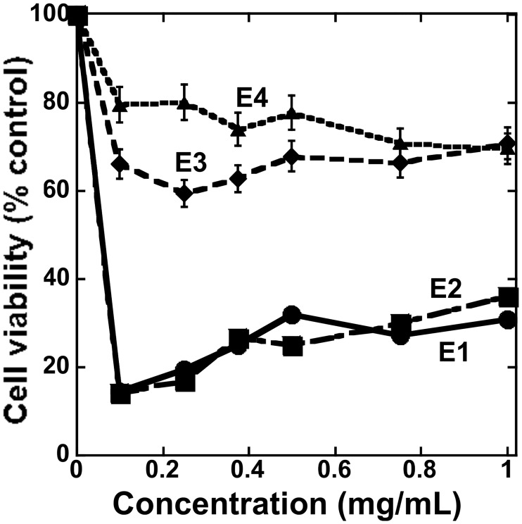
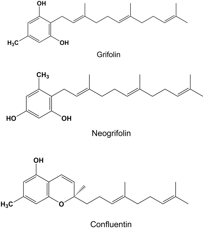
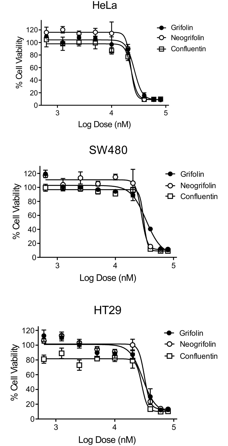
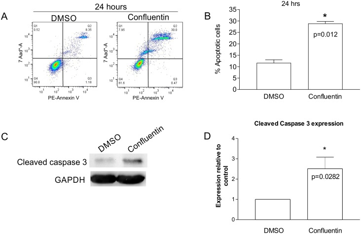
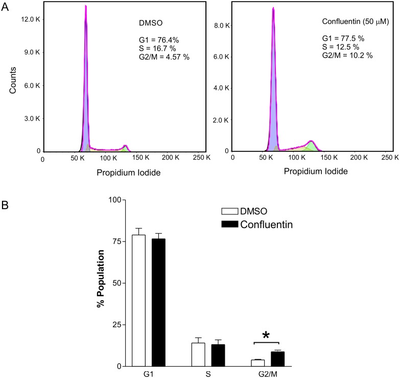
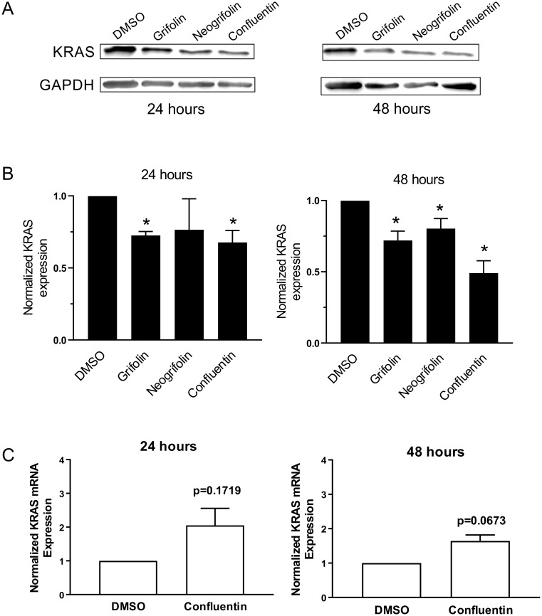
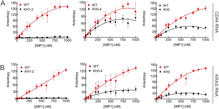
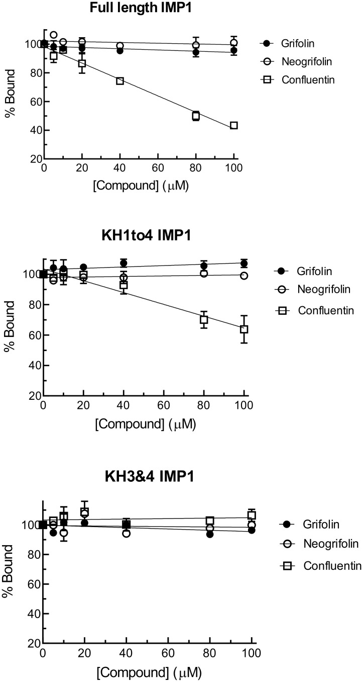
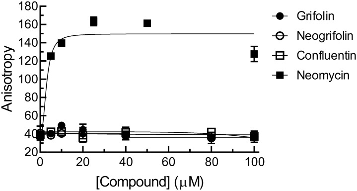
In our search for bioactive mushrooms native to British Columbia, we determined that the ethanol extracts from fruiting bodies of the terrestrial polypore Albatrellus flettii had potent anti-cell viability activity. Using bioassay-guided fractionation, mass spectrometry and nuclear magnetic resonance, we successfully isolated three known compounds (grifolin, neogrifolin and confluentin). These compounds represent the major anti-cell viability components from the ethanol extracts of A. flettii. We also identified a novel biological activity for these compounds, specifically in down-regulating KRAS expression in two human colon cancer cell lines. Relatively little is known about the anti-cell viability activity and mechanism of action of confluentin. For the first time, we show the ability of confluentin to induce apoptosis and arrest the cell cycle at the G2/M phase in SW480 human colon cancer cells. The oncogenic insulin-like growth factor 2 mRNA-binding protein 1 (IMP1) has been previously shown to regulate KRAS mRNA expression in colon cancer cells, possibly through its ability to bind to the KRAS transcript. Using a fluorescence polarization assay, we show that confluentin dose-dependently inhibits the physical interaction between KRAS RNA and full-length IMP1. The inhibition also occurs with truncated IMP1 containing the KH1 to KH4 domain (KH1to4 IMP1), but not with the di-domain KH3 and KH4 (KH3&4 IMP1). In addition, unlike the control antibiotic neomycin, grifolin, neogrifolin and confluentin do not bind to KRAS RNA. These results suggest that confluentin inhibits IMP1-KRAS RNA interaction by binding to the KH1&2 di-domains of IMP1. Since the molecular interaction between IMP1 and its target RNAs is a pre-requisite for the oncogenic function of IMP1, confluentin should be further explored as a potential inhibitor of IMP1 in vivo.


Conflict of interest statement

The authors have declared that no competing interests exist.

Figures

Fig 1
Fig 1. Dose-dependent effect of crude extracts from A. flettii on the viability of HeLa human cervical cancer cells.
Cells were treated for 48 hours with various concentrations of E1 (80% ethanol), E2 (50% methanol), E3 (water) and E4 (5% NaOH) extracts from A. flettii. The concentrations of extracts used were: 1, 0.75, 0.5, 0.25 and 0.1 mg/mL. Anti-cell viability was measured by MTT assay. Results shown represent two biological replicates. Error bars are standard deviation (S.D.).
Fig 2
Fig 2. Chemical structure of grifolin, neogrifolin and confluentin isolated from A. flettii.
Fig 3
Fig 3. Grifolin, neogrifolin and confluentin inhibited cell viability in human cancer cells.
HeLa human cervical cancer cells and human colon cancer cells (SW480 and HT29) were treated with various concentrations of grifolin, neogrifolin and confluentin for 48 hours. The concentrations of compounds used were: 80, 60, 40, 20, 10, 5, 2.5, 1.25 and 0.625 μM. Cell viability was determined using the cytotoxic MTT assay. Each dose was tested in triplicate in each experiment and the data shown is a representation of at least three independent experiments (n = 3).
Fig 4
Fig 4. Effect of confluentin on apoptosis in SW480 human colon cancer cells.
(A) SW480 cells were treated with 2% DMSO or 50 μM confluentin for 24 hours after which cells were analyzed by flow cytometry to assess for necrosis (Q1), late apoptosis (Q2), early apoptosis (Q3) and live cells (Q4). Data shown represents three independent experiments (n = 3). (B) The percentages of cells in late apoptosis (Q2) treated with 2% DMSO or 50 μM confluentin from three independent experiments were combined and shown to be statistically significant (p < 0.05). (C) As in (A), SW480 cells treated with 2% DMSO or 50 μM confluentin for 24 hours followed by isolation of cell lysates. Western blot analyses were performed and data shown is a representative from five independent experiments (n = 5). (D) The level of Cleaved Caspase-3 in confluentin-treated cells was normalized to that of GAPDH and expressed relative to DMSO-treated cells (taken as 1.0). The plotted graph is from five independent experiments. For statistical analysis, a Student’s t-test was performed (p < 0.05). Error bars are S.D.
Fig 5
Fig 5. Effect of confluentin on cell cycle in SW480 human colon cancer cells.
(A) SW480 cells treated with 2% DMSO or 50 μM confluentin were analyzed by flow cytometry for cell-cycle distribution. (B) The relative percentages of cells in different phases of cell cycle were combined from three independent experiments (n = 3) and plotted as shown. Two-way ANOVA was performed and asterisk indicates significant differences (p < 0.001). Error bars are S.D.
Fig 6
Fig 6. Effect of grifolin, neogrifolin and confluentin on KRAS expression in SW480 human colon cancer cells.
SW480 cells were treated with 2% DMSO or 50 μM grifolin, neogrifolin and confluentin for 24 and 48 hours. (A) A representative immunoblot is shown. (B) KRAS band intensity was normalized to their respective GAPDH intensity and expressed relative to the control DMSO (taken as 1.0). Data shown are mean ± SD pooled from three independent experiments (n = 3). * = p < 0.01 versus DMSO. (C) Total RNA isolated from treated cells was subjected to quantitative real-time PCR. The level of KRAS mRNA from confluentin-treated cells was normalized to that of GAPDH mRNA and expressed relative to that of DMSO-treated cells. Data shown was pooled from three biological replicates (n = 3). A student’s t-test was performed and error bars are S.D.
Fig 7
Fig 7. Establishing the fluorescence polarization method to study IMP1-RNA interaction.
Fluorescence polarization analysis of the interaction between the wild-type IMP1 and its point-mutation variants (KH1-2, KH3-4 and KH3) with the fluorescein-labeled 39-nt CD44 (A) and the 44-nt KRAS RNAs (B). Results shown are representative of three separate experiments (n = 3).
Fig 8
Fig 8. Binding curves of the full-length and truncated IMP1 to KRAS RNA as determined using fluorescence polarization.
Increasing amounts of the full-length IMP1 or truncated IMP1 (KH1to4 and KH3&4) were incubated with 44-nt fluorescein-labeled KRAS RNA at 37°C for 30 min. Results shown represent three replicate experiments (n = 3).
Fig 9
Fig 9. Effect of grifolin, neogrifolin and confluentin on the physical interaction between KRAS RNA and IMP1 protein.
Recombinant IMP1 protein (300 nM) was incubated with fluorescently-labeled KRAS RNA in the absence or presence of various concentrations of grifolin, neogrifolin or confluentin. The concentrations of compounds used were: 100, 80, 40, 20, 10 and 5 μM. The three types of recombinant IMP1 proteins used were: full-length, KH1to4 and KH3&4. Data shown are mean ± S.D. pooled from three independent experiments (n = 3).
Fig 10
Fig 10. Grifolin (1), neogrifolin (2) and confluentin (3) do not bind to KRAS RNA.
Increasing concentrations of grifolin, neogrifolin and confluentin were incubated with the 44-nt fluorescein-labeled KRAS RNA at 37°C for 30 min. Fluorescence anisotropy units were then measured. Neomycin was used as the positive control. The results shown represent three replicate experiments (n = 3).

References

    1. Smith A, Javed S, Barad A, Myhre V, Li WM, Reimer K, et al. Growth-inhibitory and immunomodulatory activities of wild mushrooms from north-central British Columbia (Canada). Int J Med Mushrooms. 2017;19: 485–497. 10.1615/IntJMedMushrooms.v19.i6.10 - DOI - PubMed
    1. Deo G, Khatra J, Buttar S, Li WM, Tackaberry LE, Massicotte H.B, et al. Anti-proliferative, immuno-stimulatory and anti-inflammatory activities of extracts derived from mushrooms collected in Haida Gwaii, British Columbia, Canada. Int J Med Mushrooms. 2019;21: 629–643. 10.1615/IntJMedMushrooms.2019031193 - DOI - PubMed
    1. Ginns J. The taxonomy and distribution of rare or uncommon species of Albatrellus in western North America. Can J Bot. 1997;75: 261–273.
    1. Nukata M, Hashimoto T, Yamamoto I, Iwasaki N, Tanaka M, Asakawa Y. Neogrifolin derivatives possessing anti-oxidative activity from the mushroom Albatrellus ovinus. Phytochem. 2002;59: 731–737. - PubMed
    1. Liu L-Y, Li Z-H, Ding Z-H, Dong Z-J, Li G-T, Li Y, et al. Meroterpenoid pigments from the basidiomycete Albatrellus ovinus. J Nat Prod. 2013;76: 79–84. 10.1021/np300751m - DOI - PubMed

Publication types

MeSH terms