. 2020 Jul 30;136(5):627-639.
doi: 10.1182/blood.2019003442.

Platelets docking to VWF prevent leaks during leukocyte extravasation by stimulating Tie-2

Affiliations

Platelets docking to VWF prevent leaks during leukocyte extravasation by stimulating Tie-2

Laura J Braun et al. Blood. .

Abstract

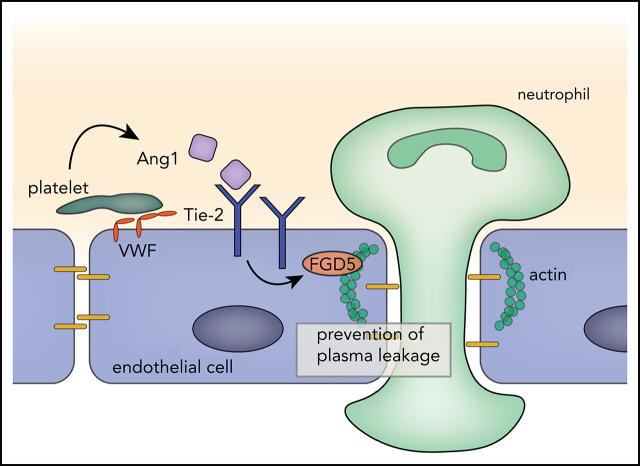
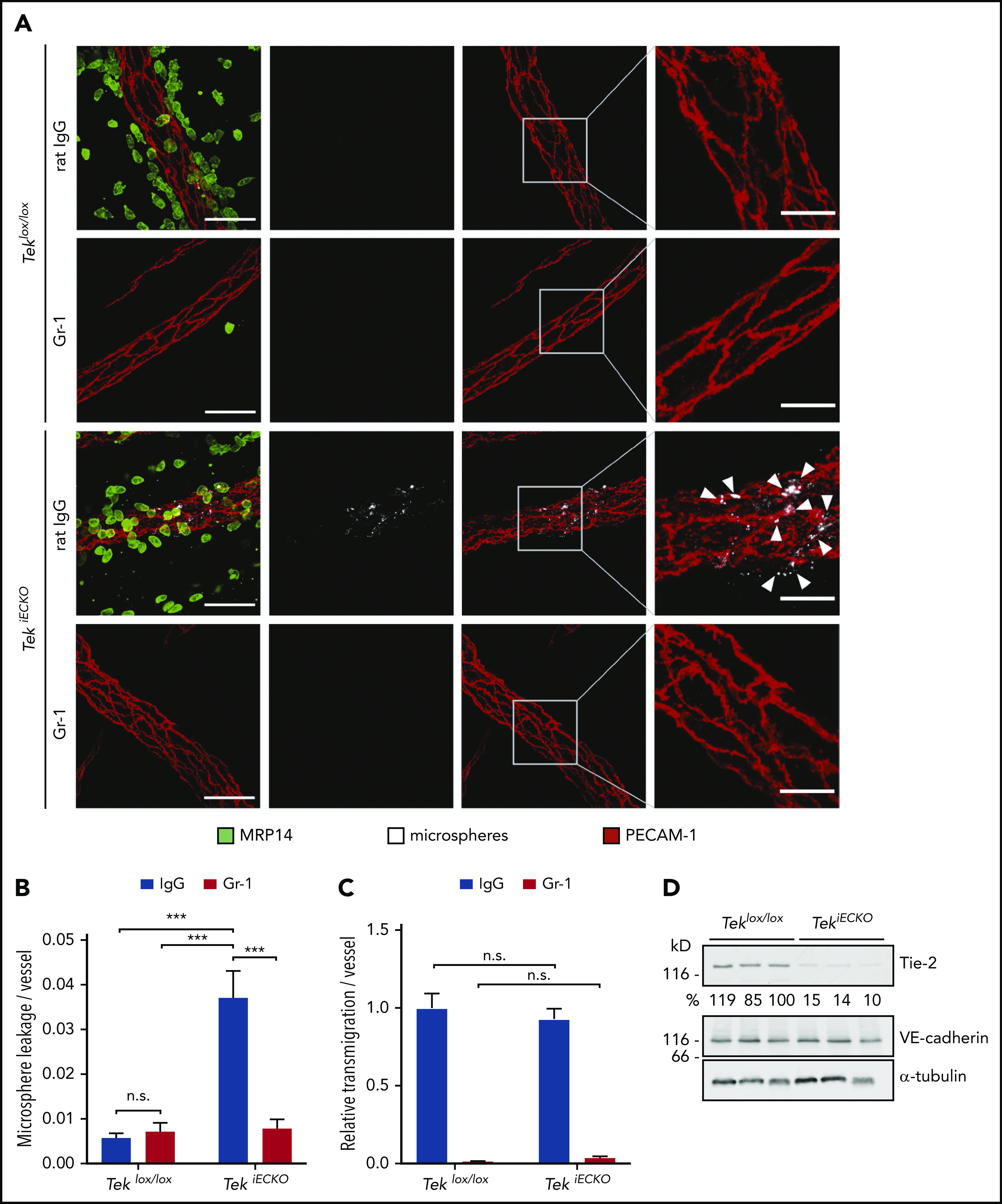
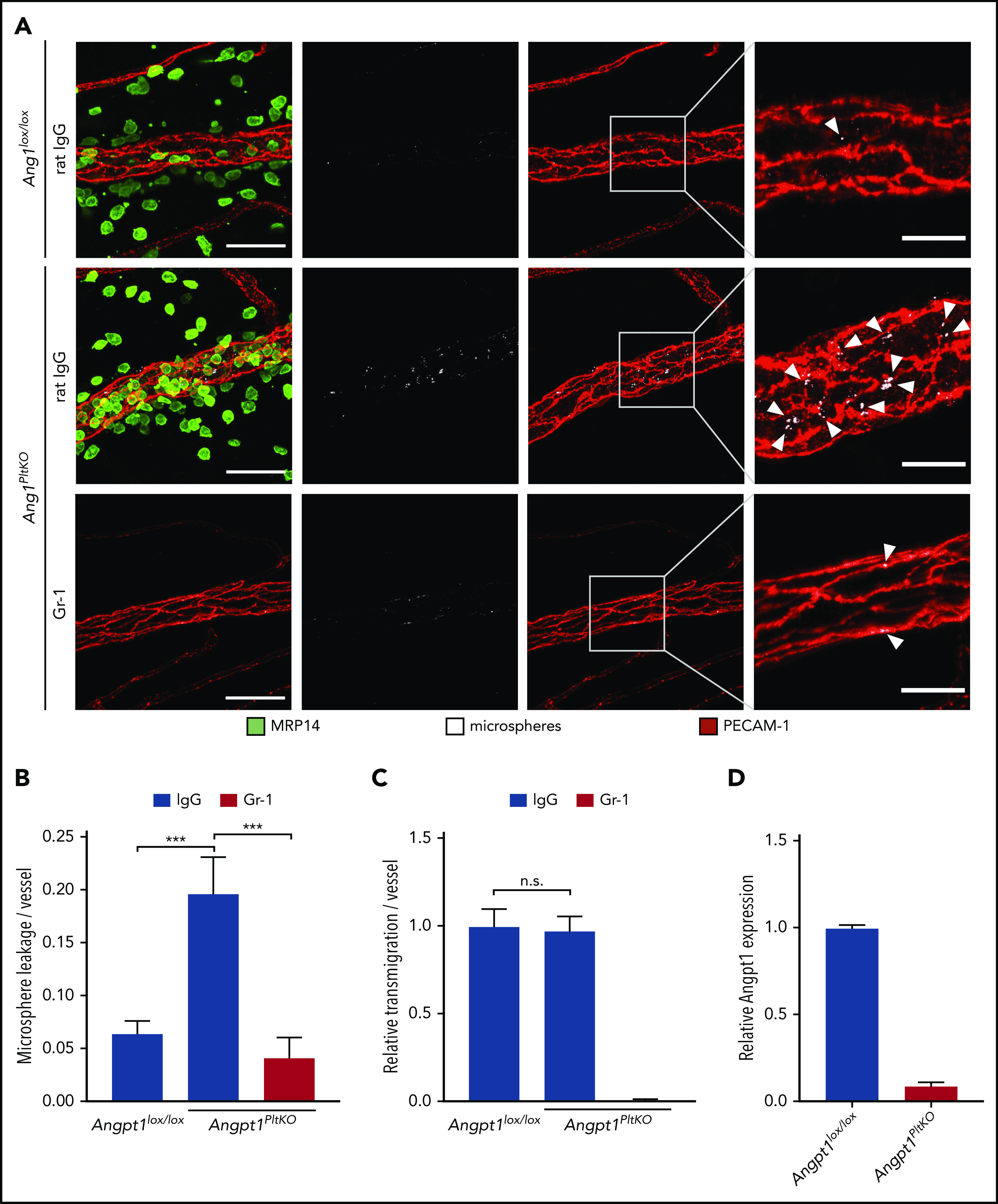
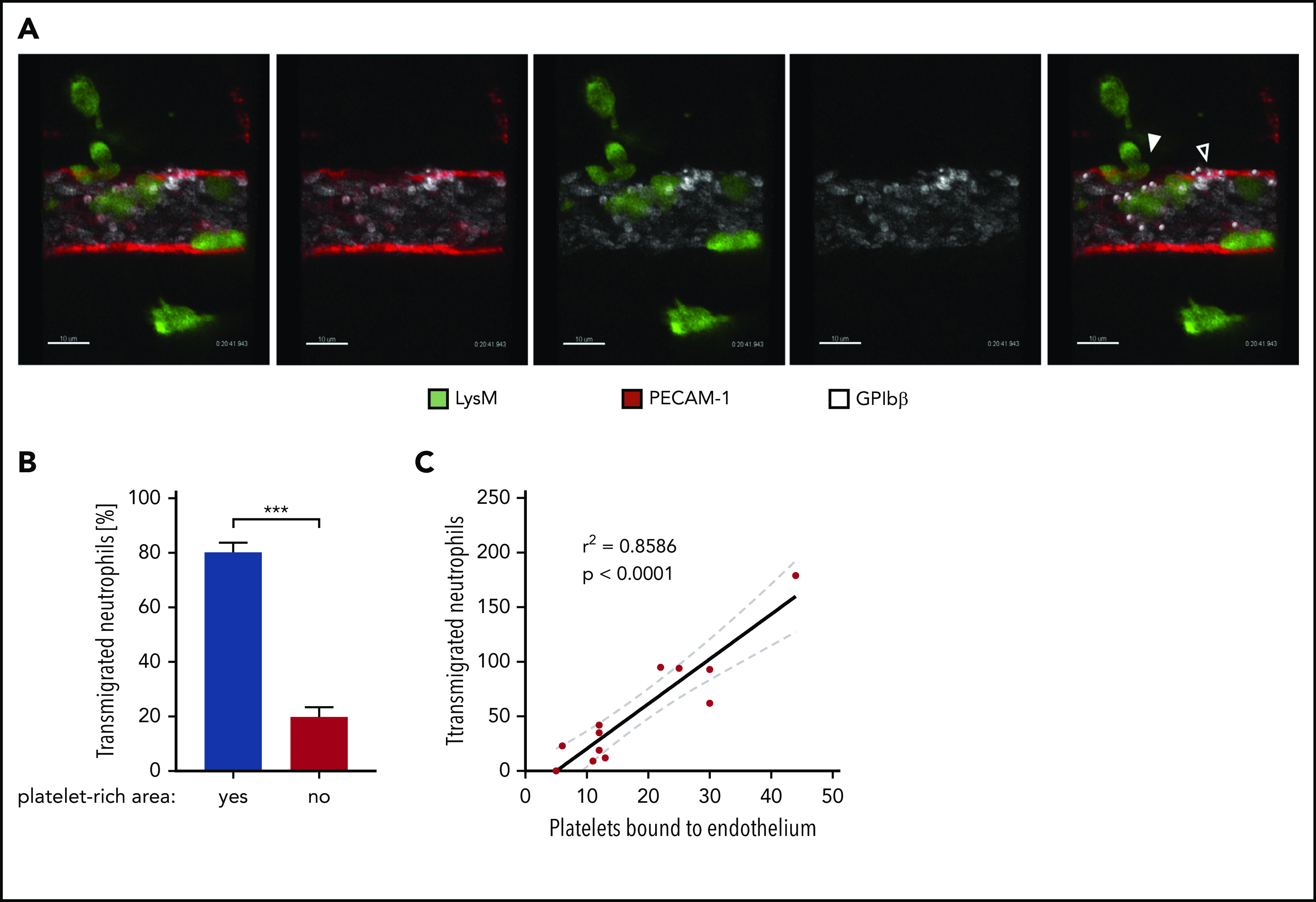
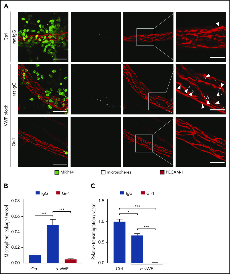
Neutrophil extravasation requires opening of the endothelial barrier but does not necessarily cause plasma leakage. Leaks are prevented by contractile actin filaments surrounding the diapedesis pore, keeping this opening tightly closed around the transmigrating neutrophils. We have identified the receptor system that is responsible for this. We show that silencing, or gene inactivation, of endothelial Tie-2 results in leak formation in postcapillary venules of the inflamed cremaster muscle at sites of neutrophil extravasation, as visualized by fluorescent microspheres. Leakage was dependent on neutrophil extravasation, because it was absent upon neutrophil depletion. We identified the Cdc42 GTPase exchange factor FGD5 as a downstream target of Tie-2 that is essential for leakage prevention during neutrophil extravasation. Looking for the Tie-2 agonist and its source, we found that platelet-derived angiopoietin-1 (Angpt1) was required to prevent neutrophil-induced leaks. Intriguingly, blocking von Willebrand factor (VWF) resulted in vascular leaks during transmigration, indicating that platelets interacting with endothelial VWF activate Tie-2 by secreting Angpt1, thereby preventing diapedesis-induced leakiness.


Conflict of interest statement

Conflict-of-interest disclosure: The authors declare no competing financial interests.

Figures

None
Graphical abstract
Figure 1.
Figure 1.
FGD5 is necessary to prevent neutrophil-induced leakiness in vitro and in vivo. (A) Paracellular permeability for 40 kDa FITC-dextran was determined for TNF-α–stimulated HUVEC that were pretreated with control (Ctrl) or FGD5 siRNA in the presence or absence of human neutrophils freshly isolated from whole blood (as indicated). Values were normalized to Ctrl siRNA monolayers without PMNs. (B) Relative transmigration of human PMNs across HUVEC monolayers treated with Ctrl or FGD5 siRNA. (C) HUVEC transfected with Ctrl or FGD5-targeting siRNA were immunoblotted for FGD5 and α-tubulin. (D) Mice were injected intrascrotally with FGD5 or Ctrl siRNA and received anti–Gr-1 antibodies or control IgG intraperitoneally the following day to deplete neutrophils. Twenty-four hours later, mice were stimulated with IL-1β for 3 hours before fluorescent microspheres were injected IV, and mice were euthanized 5 minutes later. Whole mounts of the cremaster muscle were stained with antibodies against PECAM-1 and MRP14. Arrowheads indicate microsphere leakage. Scale bars, 40 µm (left panels), 15 µm (right panels). Quantification of microspheres (E) and extravasated neutrophils (F) per vessel area. (G) Total lung lysates of mice treated with Ctrl siRNA or FGD5 siRNA were immunoblotted for the indicated antigens. Data are mean ± SEM. Results are representative of (C,D,G) or pooled from 4 independent experiments (A-B) or pooled from 4 independent experiments, with a total of 35 to 40 vessels analyzed per condition (E-F). *P < .05, **P < .01, ***P < .001, Student t test (B), 2-way ANOVA (A,E-F). n.s., not significant.
Figure 2.
Figure 2.
Tie-2 siRNA promotes neutrophil-dependent microsphere leakage from inflamed cremaster vessels. (A) Mice received control (Ctrl) siRNA or Tie-2 siRNA intrascrotally, 24 hours later they received intraperitoneal anti–Gr-1 antibodies or control IgG, and 24 hours thereafter they received IL-1β intrascrotally, followed by IV injection of fluorescent microspheres 3 hours later; mice were euthanized 5 minutes later. Whole mounts of the cremaster muscle were stained for PECAM-1 and MRP14. Arrowheads indicate microphere leakage. Scale bars, 40 µm (left panels), 15 µm (right panels). (B) Microsphere leakage and (C) relative neutrophil extravasation per vessel from experiments as in (A). (D) Total lung lysates from Ctrl siRNA–treated or Tie-2 siRNA–treated mice were analyzed by immunoblot for expression of Tie-2, VE-cadherin, and α-tubulin. Results are representative of (A,D) or pooled from (B-C) 3 independent experiments, with a total of 30 vessels analyzed per condition. Data are mean ± SEM. ***P < .001, 2-way ANOVA. n.s., not significant.
Figure 3.
Figure 3.
Conditional gene inactivation of Tek in mice. (A-B) Teklox/lox and TekiECKO mice were injected intraperitoneally with tamoxifen on 5 consecutive days and analyzed 3 days later. (A) Total lysates of lung, heart and skin were immunoblotted for Tie-2, VE-cadherin and α-tubulin. (B) Cremaster whole mounts were stained for Tie-2 and PECAM-1. Scale bars, 50 µm.
Figure 4.
Figure 4.
Conditional inactivation of Tie-2 in endothelial cells evokes neutrophil-dependent leakiness of microspheres. (A) Teklox/lox and TekiECKO mice received intraperitoneal injections of anti–Gr-1 or control IgG antibodies and were stimulated 24 hours later for 3 hours with IL-1β. Fluorescent microspheres were injected IV for 5 minutes, and cremaster whole mounts were stained for PECAM-1 and MRP14. Arrowheads indicate microsphere leakage. Scale bars, 40 µm (left panels), 15 µm (right panels). Quantification of microsphere leakage (B) and relative neutrophil extravasation (C) per vessel. (D) Total lung lysates of Teklox/lox and TekiECKO mice were analyzed by immunoblot for the indicated antigens. Results are representative of (A,D) or pooled from (B-C) 4 independent experiments, with 40 or 41 vessels analyzed. Data are mean ± SEM. ***P < .001, 2-way ANOVA. n.s., not significant.
Figure 5.
Figure 5.
Platelet-derived Angpt1 prevents neutrophil-induced plasma leakage. (A-C) Angpt1lox/lox and Angpt1PltKO mice were injected intraperitoneally with anti–Gr-1 or control IgG antibodies to deplete neutrophils and were stimulated intrascrotally 24 hours later with IL-1β. Four hours later, fluorescent microspheres were injected IV, and cremaster whole mounts were prepared and stained for PECAM-1 and MRP14 5 minutes later. Arrowheads indicate microsphere leakage. Scale bars, 40 µm (left panels), 15 µm (right panels). Microsphere leakage (B) and transmigrated neutrophils (C) per vessel were quantified. (D) Lysates of platelets isolated from Angpt1lox/lox or Angpt1PltKO mice were submitted to an Angpt1 enzyme-linked immunosorbent assay (Bosterbio), according to the manufacturer’s instructions, to measure Angpt1 content. Results are representative of 5 (A) or are pooled from 3 (D) or 5 (B-C) independent experiments, with 50 vessels analyzed. Data are mean ± SEM. ***P < .001, 1-way ANOVA. n.s., not significant.
Figure 6.
Figure 6.
Preferential extravasation of neutrophils in platelet-rich areas of cremasteric venules. (A) Confocal intravital microscopy of cremasteric venules in LysM-eGFP mice that were injected intrascrotally with IL-1β. Endothelial cells and platelets were labeled through IV injection of fluorophore-coupled anti–PECAM-1 and anti–GPIbβ antibodies, respectively. Mice were anaesthetized, the cremaster muscle was prepared for intravital imaging, and videos were taken for 1 to 1.5 hours. White dots mark platelets bound to the endothelium, the filled arrowhead marks areas of neutrophil extravasation, and the open arrowhead marks areas of extravasation (supplemental Video 1). Scale bars, 10 µm. The timestamp of the video is 0:20:41:943. (B) Quantification of neutrophil transmigration in platelet-rich and platelet-poor areas of cremasteric venules from 10 videos shown as the percentage of total transmigrated neutrophils. Data are mean ± SEM. (C) Correlation analysis (Pearson) and linear regression of transmigrated neutrophils and endothelium-bound platelets in videos, as described in (A). Results are representative of (A) or pooled from (B-C) 10 videos taken of 9 mice. ***P < .001, Mann-Whitney rank test.
Figure 7.
Figure 7.
Blockade of endothelial VWF prevents microsphere leakage during neutrophil transmigration. (A) C57BL/6 mice received anti–Gr-1 antibodies or control (Ctrl) IgG and were injected IV with VWF-blocking or control antibodies 24 hours later. Five minutes later, local inflammation was induced by intrascrotal IL-1β injection. Fluorescent microspheres were injected IV 4 hours later, and cremaster whole mounts were prepared and stained for PECAM-1 and MRP14 5 minutes later. Arrowheads indicate microsphere leakage. Scale bars, 40 µm (left panels), 15 µm (right panels). Microsphere leakage (B) and neutrophil extravasation (C) in experiments as depicted in (A) were quantified and normalized to vessel surface. Results are representative of (A) or pooled from (B-C) 5 independent experiments, with 50 vessels analyzed. Data are mean ± SEM. *P < .05, ***P < .001, 1-way ANOVA.

Comment in

References

    1. Carden DL, Smith JK, Korthuis RJ. Neutrophil-mediated microvascular dysfunction in postischemic canine skeletal muscle. Role of granulocyte adherence. Circ Res. 1990;66(5):1436-1444. - PubMed
    1. Kubes P, Suzuki M, Granger DN. Modulation of PAF-induced leukocyte adherence and increased microvascular permeability. Am J Physiol. 1990;259(5 Pt 1):G859-G864. - PubMed
    1. Sumagin R, Lomakina E, Sarelius IH. Leukocyte-endothelial cell interactions are linked to vascular permeability via ICAM-1-mediated signaling. Am J Physiol Heart Circ Physiol. 2008;295(3):H969-H977. - PMC - PubMed
    1. DiStasi MR, Ley K. Opening the flood-gates: how neutrophil-endothelial interactions regulate permeability. Trends Immunol. 2009;30(11):547-556. - PMC - PubMed
    1. Finsterbusch M, Voisin MB, Beyrau M, Williams TJ, Nourshargh S. Neutrophils recruited by chemoattractants in vivo induce microvascular plasma protein leakage through secretion of TNF. J Exp Med. 2014;211(7):1307-1314. - PMC - PubMed

Publication types