. 2020 Jul;246(1):69-78.
doi: 10.1530/JOE-19-0535.

Genetic deletion of Urocortin 3 does not prevent functional maturation of beta cells

Affiliations

Genetic deletion of Urocortin 3 does not prevent functional maturation of beta cells

Jessica L Huang et al. J Endocrinol. 2020 Jul.

Abstract

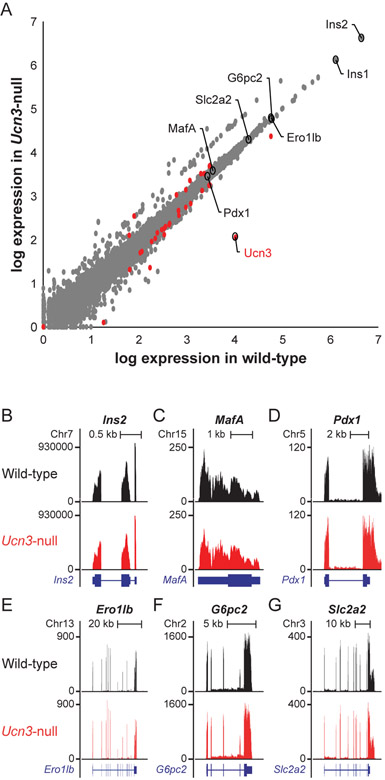
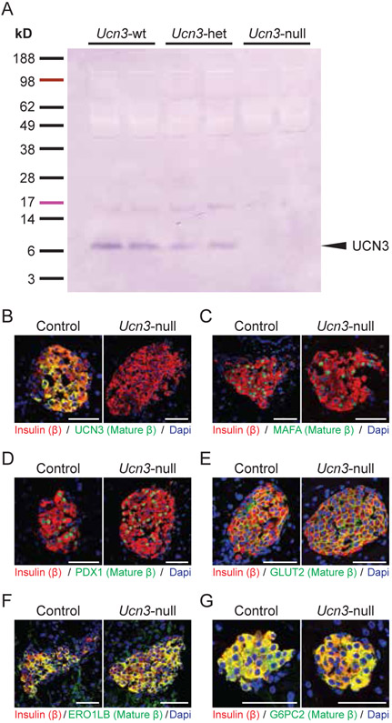
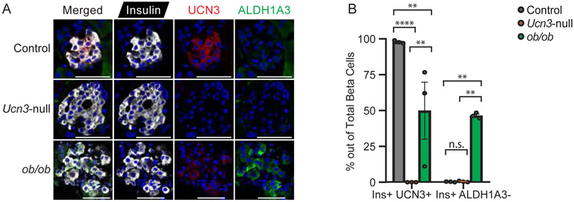
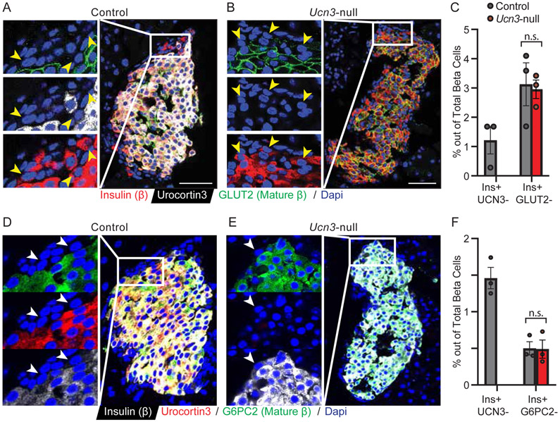
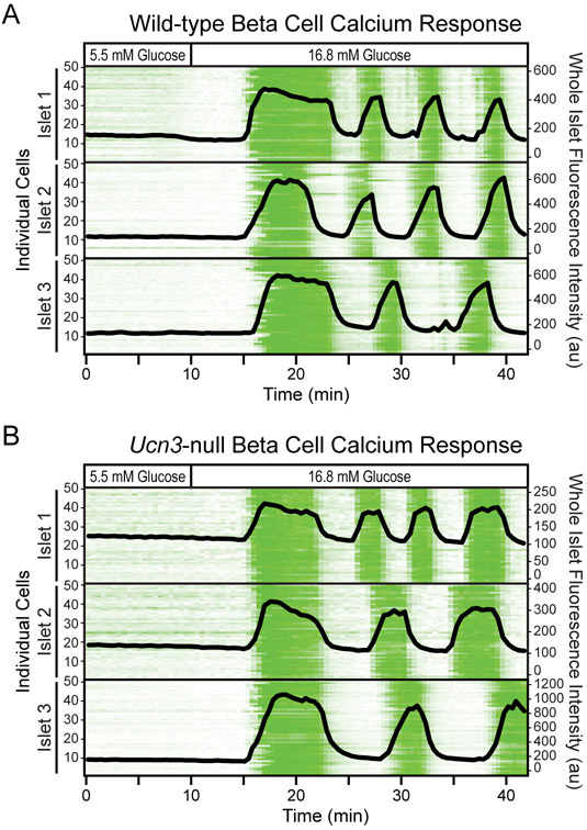
There is great interest in generating functionally mature beta cells from stem cells, as loss of functional beta cell mass contributes to the pathophysiology of diabetes. Identifying markers of beta cell maturity is therefore very helpful for distinguishing stem cells that have been successfully differentiated into fully mature beta cells from stem cells that did not. Urocortin 3 (UCN3) is a peptide hormone whose expression is associated with the acquisition of functional maturity in beta cells. The onset of its expression occurs after other beta cell maturity markers are already expressed and its loss marks the beginning of beta cell dedifferentiation. Its expression pattern is therefore tightly correlated with beta cell maturity. While this makes UCN3 an excellent marker of beta cell maturity, it is not established whether UCN3 is required for beta cell maturation. Here, we compared gene expression and function of beta cells from Ucn3-null mice relative to WT mice to determine whether beta cells are functionally mature in the absence of UCN3. Our results show that genetic deletion of Ucn3 does not cause a loss of beta cell maturity or an increase in beta cell dedifferentiation. Furthermore, virgin beta cells, first identified as insulin-expressing, UCN3-negative beta cells, can still be detected at the islet periphery in Ucn3-null mice. Beta cells from Ucn3-null mice also exhibit normal calcium response when exposed to high glucose. Collectively, these observations indicate that UCN3 is an excellent mature beta cell marker that is nevertheless not necessary for beta cell maturation.

Keywords: GLUT2; UCN3; Urocortin 3; beta cell maturity; pancreatic beta cell.


Conflict of interest statement

Declaration of Interest

The authors declare no conflicts of interest.

Figures

Figure 1.
Figure 1.. Genes encoding beta cell maturity markers are not differentially regulated between Ucn3-null mice and wild-type littermates.
(A) Volcano plot showing global gene expression in wild-type vs Ucn3-null mice. Differentially expressed genes are denoted as red circles. Select markers of beta cell maturity are circled and labeled. Ucn3 is depleted in Ucn3-null mice, confirming knockout, while other beta cell maturity markers are not affected at the transcript level. (B-G) Visualization of relative reads of different transcripts in Ucn3-null mice (red) compared to wild-type (black). (B) Ins2, (C) MafA, (D) Pdx1, (E) Ero1lb, (F) G6pc2, (G) Slc2a2.
Figure 2.
Figure 2.. UCN3 is not necessary for beta cells to express maturity markers.
(A,B) Western blot and immunofluorescence confirm the loss of UCN3 in Ucn3-null mice. (B-G) Co-localization of insulin and select beta cell maturity markers in islets of Ucn3-null mice and littermate controls. Insulin is stained in red, the marker of interest is in green, and nuclei stained by DAPI are in blue. (B) UCN3, (C) MAFA, (D) PDX1, (E) ERO1LB, (F) G6PC2, (G) GLUT2. Scale bars indicate 50 μm.
Figure 3.
Figure 3.. The absence of UCN3 does not lead to beta cell dedifferentiation.
(A) Immunofluorescence of insulin, UCN3, and ALDH1A3 in islets from wild-type, Ucn3-null, and ob/ob mice. Control mice co-express insulin and UCN3 in beta cells, but express little to no ALDH1A3. In contrast, more insulin-positive beta cells in ob/ob islets express ALDH1A3 and exhibit weaker immunoreactivity of UCN3. Ucn3-null mice, like their control littermates, express little to no ALDH1A3 in beta cells but lack expression of UCN3. Scale bars indicate 50 μm. (B) Quantification of insulin-positive, UCN3-positive and insulin-positive, ALDH1A3-positive cells in control, Ucn3-null, and ob/ob mice, as percentage of total beta cells. n = 3 animals per group, at least 10 islets and 500 cells counted per animal. Error bars reflect SEM. **p<0.01, ****p<0.001
Figure 4.
Figure 4.. Virgin beta cells are maintained at the islet periphery in the absence of UCN3.
(A-B) Immunofluorescence of insulin, UCN3, and GLUT2 in islets from (A) control and (B) Ucn3-null mice. Insulin and UCN3 co-localize in beta cells, while GLUT2 is expressed on the membrane of the beta cells. Arrows point to virgin beta cells, which are insulin-positive but UCN3-negative and GLUT2-negative in control mice and insulin-positive but GLUT2-negative in Ucn3-null mice. Scale bars indicate 50 μm. (C) Quantification of insulin-positive, UCN3-negative cells in control mice and insulin-positive, csGLUT2-negative cells in control and Ucn3-null mice, as percentage of total beta cells. (D-E) Immunofluorescence of insulin, UCN3, and G6PC2 in islets from (A) control and (B) Ucn3-null mice. Insulin, UCN3, and G6PC2 are co-expressed in the majority of beta cells in control mice. Arrows point to virgin beta cells that are insulin-positive but UCN3- and G6PC2-negative. (F) Quantification of insulin-positive, UCN3-negative cells in control mice and insulin-positive, G6PC2-negative cells in control and Ucn3-null mice. For both C and F, there is no quantification for insulin+ UCN3- cells in the Ucn3-null mice as all cells are UCN3- and thus not representative of the virgin beta cell population. n = 3 animals per group, at least 10 islets and 500 cells counted per animal. Error bars reflect SEM.
Figure 5.
Figure 5.. Beta cell calcium response remains unaltered in mice that do not express UCN3.
Calcium responses of several intact islets were obtained simultaneously over time. All islets expressed the calcium sensor GCaMP6s selectively in beta cells (MIP-Cre/ERT x lsl-GCaMP6s) with or without Ucn3-null in the background. The trace began with low glucose and was switched to high glucose at the indicated timepoint. The calcium traces of beta cells within 3 representative islets from (A) wild-type mice and (B) Ucn3-null mice are shown as intensity plots. Each line represents the activity of an individual beta cell over time, and green represents an increase in fluorescence intensity. A line graph of the fluorescence intensity at the whole islet level is overlaid over each intensity plot.

References

    1. Artner I, Hang Y, Mazur M, Yamamoto T, Guo M, Lindner J, Magnuson MA & Stein R 2010. MafA and MafB regulate genes critical to β-cells in a unique temporal manner. Diabetes 59 2530–2539. (doi:10.2337/db10-0190) - DOI - PMC - PubMed
    1. Beamish CA, Strutt BJ, Arany EJ & Hill DJ 2016. Insulin-positive, Glut2-low cells present within mouse pancreas exhibit lineage plasticity and are enriched within extra-islet endocrine cell clusters. Islets 8 65–82. (doi:10.1080/19382014.2016.1162367) - DOI - PMC - PubMed
    1. Bergsten P, Grapengiesser E, Gylfe E, Tengholm A & Hellman B 1994. Synchronous oscillations of cytoplasmic Ca2+ and insulin release in glucose-stimulated pancreatic islets. Journal of Biological Chemistry 269 8749–8753. - PubMed
    1. Blum B, Hrvatin S, Schuetz C, Bonal C, Rezania A & Melton DA 2012. Functional beta-cell maturation is marked by an increased glucose threshold and by expression of urocortin 3. Nature Biotechnology 30 261–264. (doi:10.1038/nbt.2141) - DOI - PMC - PubMed
    1. Blum B, Roose AN, Barrandon O, Maehr R, Arvanites AC, Davidow LS, Davis JC, Peterson QP, Rubin LL & Melton DA 2014. Reversal of β cell de-differentiation by a small molecule inhibitor of the TGFβ pathway. ELife 3 e02809. (doi:10.7554/eLife.02809) - DOI - PMC - PubMed

Publication types

MeSH terms