. 2020 May 1;8(5):659.
doi: 10.3390/microorganisms8050659.

Virulence Characteristics of mecA-Positive Multidrug-Resistant Clinical Coagulase-Negative Staphylococci

Affiliations

Virulence Characteristics of mecA-Positive Multidrug-Resistant Clinical Coagulase-Negative Staphylococci

Jung-Whan Chon et al. Microorganisms. .

Abstract

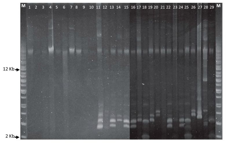
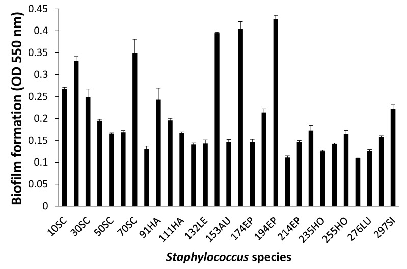
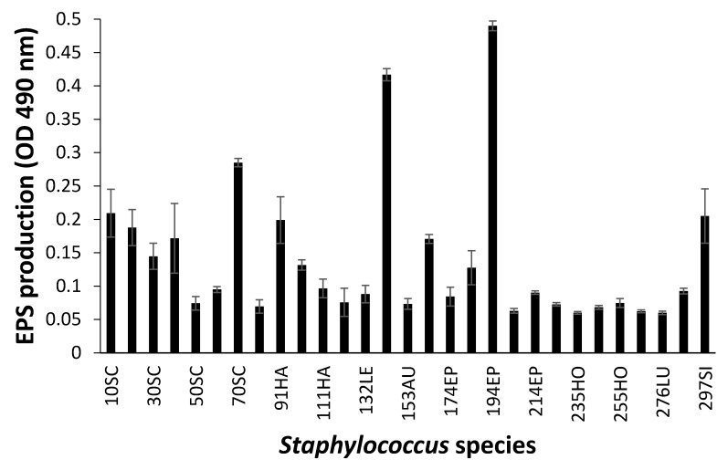
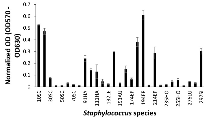
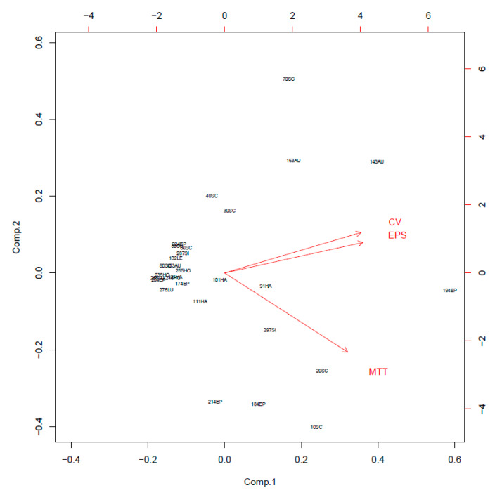
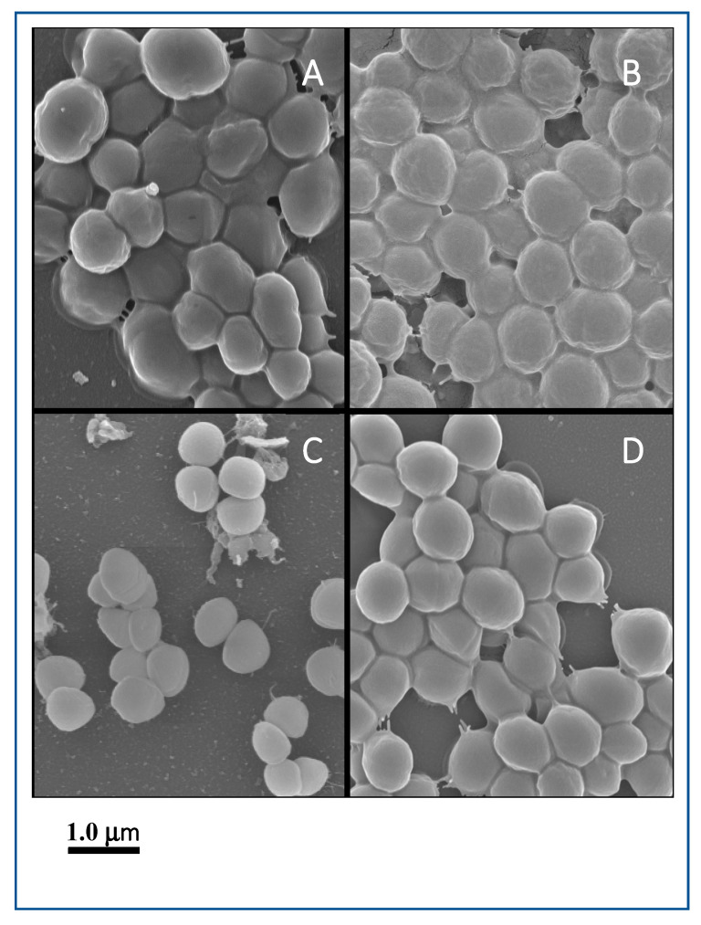
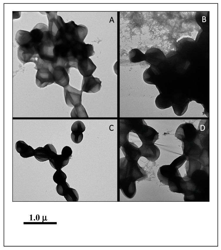
Coagulase-negative staphylococci (CoNS) are an important group of opportunistic pathogenic microorganisms that cause infections in hospital settings and are generally resistant to many antimicrobial agents. We report on phenotypic and genotypic virulence characteristics of a select group of clinical, mecA-positive (encoding penicillin-binding protein 2a) CoNS isolates. All CoNS were resistant to two or more antimicrobials with S. epidermidis strain 214EP, showing resistance to fifteen of the sixteen antimicrobial agents tested. Aminoglycoside-resistance genes were the ones most commonly detected. The presence of megaplasmids containing both horizontal gene transfer and antimicrobial resistance genetic determinants indicates that CoNS may disseminate antibiotic resistance to other bacteria. Staphylococcus sciuri species produced six virulence enzymes, including a DNase, gelatinase, lipase, phosphatase, and protease that are suspected to degrade tissues into nutrients for bacterial growth and contribute to the pathogenicity of CoNS. The PCR assay for the detection of biofilm-associated genes found the eno (encoding laminin-binding protein) gene in all isolates. Measurement of their biofilm-forming ability and Spearman's rank correlation coefficient analyses revealed that the results of crystal violet (CV) and extracellular polymeric substances (EPS) assays were significantly correlated (ρ = 0.9153, P = 3.612e-12). The presence of virulence factors, biofilm-formation capability, extracellular enzymes, multidrug resistance, and gene transfer markers in mecA-positive CoNS clinical strains used in this study makes them powerful opportunistic pathogens. The study also warrants a careful evaluation of nosocomial infections caused by CoNS and may be useful in studying the mechanism of virulence and factors associated with their pathogenicity in vivo and developing effective strategies for mitigation.

Keywords: coagulase-negative staphylococci; multidrug-resistant; virulence.


Conflict of interest statement

The authors declare no conflict of interest.

Figures

Figure 1
Figure 1
Plasmid DNA profiles of Staphylococcus species. Agarose gel electrophoresis (0.8%) was run at 100 V for 4 h and a supercoiled DNA ladder from Agilent Technologies was used as a molecular size marker. First and last lanes are supercoiled plasmid DNA ladders. Staphylococcus strains in lanes 1-29—10SC, 20SC, 30SC, 40SC, 50SC, 60SC, 70SC, 80SC, 91HA, 101HA, 111HA, 121HA, 132LE, 143AU, 153AU, 163AU, 174EP, 184EP, 194EP, 204EP, 214EP, 224EP, 235HO, 245HO, 255HO, 266LU, 276LU, 287SI, and 297SI.
Figure 2
Figure 2
Biofilm formation of Staphylococcus species shown by crystal violet (CV) staining. Experiments were run in triplicate and each bar represents the mean ± standard deviation from the mean.
Figure 3
Figure 3
Biofilm formation of Staphylococcus species shown by extracellular polymeric substances (EPS) assay. Experiments were run in triplicate and each bar represents the mean ± standard deviation from the mean.
Figure 4
Figure 4
Biofilm formation of Staphylococcus species shown by 3-(4,5-dimethyl-2-thiazolyl)-2,5-diphenyl-2H-tetrazolium bromide (MTT) assay. Experiments were run in triplicate and each bar represents the mean ± standard deviation from the mean.
Figure 5
Figure 5
Principal component analysis (PCA) of crystal violet (CV), extracellular polymeric substances (EPS), and 3-(4,5-dimethyl-2-thiazolyl)-2,5-diphenyl-2H-tetrazolium bromide (MTT) assays.
Figure 6
Figure 6
Field emission scanning electron microscopy (FESEM) images of biofilm. The scale bar in all the images corresponds to 1.0 μm. (A): S. sciuri strain 10SC, (B): S. sciuri strain 70SC, (C): S. lugdunensis strain 266LU, (D): S. simulans strain 297SI.
Figure 7
Figure 7
NS-TEM images of biofilm. The scale bar in all the images corresponds to 1.0 μm. (A): S. sciuri strain 10SC, (B): S. sciuri strain 70SC, (C): S. lugdunensis strain 266LU, (D): S. simulans strain 297SI.

Similar articles

Cited by

References

    1. Becker K., Heilmann C., Peters G. Coagulase-negative staphylococci. Clin. Microbiol. Rev. 2014;27:870–926. doi: 10.1128/CMR.00109-13. - DOI - PMC - PubMed
    1. Couto I., Pereira S., Miragaia M., Sanches I.S., de Lencastre H. Identification of clinical staphylococcal isolates from humans by internal transcribed spacer PCR. J. Clin. Microbiol. 2001;39:3099–3103. doi: 10.1128/JCM.39.9.3099-3103.2001. - DOI - PMC - PubMed
    1. Piette A., Verschraegen G. Role of coagulase-negative staphylococci in human disease. Vet. Microbiol. 2009;134:45–54. doi: 10.1016/j.vetmic.2008.09.009. - DOI - PubMed
    1. Szczuka E., Jablonska L., Kaznowski A. Coagulase-negative staphylococci: Pathogenesis, occurrence of antibiotic resistance genes and in vitro effects of antimicrobial agents on biofilm-growing bacteria. J. Med. Microbiol. 2016;65:1405–1413. doi: 10.1099/jmm.0.000372. - DOI - PubMed
    1. Vautor E., Abadie G., Pont A., Thiery R. Evaluation of the presence of the bap gene in Staphylococcus aureus isolates recovered from human and animals species. Vet. Microbiol. 2008;127:407–411. doi: 10.1016/j.vetmic.2007.08.018. - DOI - PubMed

LinkOut - more resources