

Diffusion-Weighted Magnetic Resonance Imaging as a Noninvasive Parameter for Differentiating Benign and Malignant Intraperitoneal Collections
- PMID: 32369983
- PMCID: PMC7279298
- DOI: 10.3390/medicina56050217
Diffusion-Weighted Magnetic Resonance Imaging as a Noninvasive Parameter for Differentiating Benign and Malignant Intraperitoneal Collections
Abstract
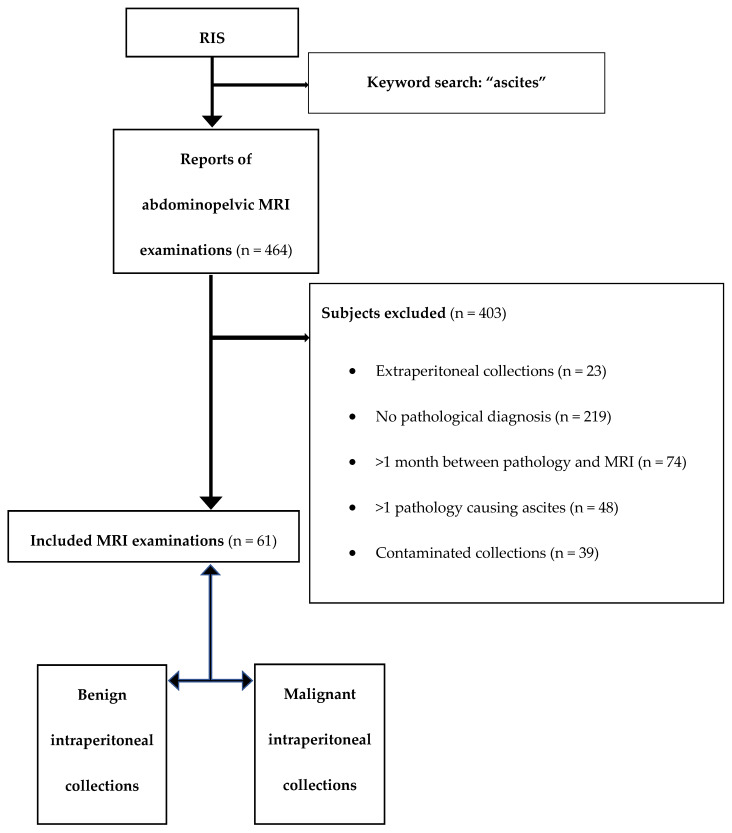
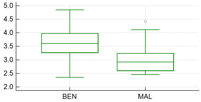
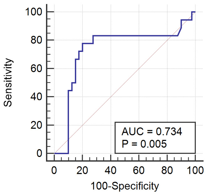
Background and Objective: The imaging differentiation of benign from malignant intraperitoneal collections (IPCs) relies on the tumoral morphological modifications of the peritoneum, which are not always advocating for malignancy. We aimed to assess ascitic fluid with the apparent diffusion coefficient (ADC) to determine non-invasive, stand-alone, differentiation criteria for benign and malignant intraperitoneal effusions. Materials and Methods: Sixty-one patients with known IPCs who underwent magnetic resonance examinations for reasons such as tumor staging, undetermined abdominal mass and disease follow up were retrospectively included in this study. All subjects had a final diagnosis of the fluid based on pathological examinations, which were divided into benign (n = 37) and malignant (n = 24) IPCs groups. ADC values were measured separately by two radiologists, and the average values were used for comparing the two groups by consuming the independent samples t-test. The receiver operating characteristic analysis was performed to test the ADC values' diagnostic ability to distinguish malignant from benign collections. Results: The differentiation between benign and malignant IPCs based on ADC values was statistically significant (p = 0.0034). The mean ADC values were higher for the benign (3.543 × 10⁻³ mm2/s) than for the malignant group (3.057 × 10⁻³ mm2/s). The optimum ADC cutoff point for the diagnosis of malignant ascites was <3.241 × 10⁻³ mm2/s, with a sensitivity of 77.78% and a specificity of 80%. Conclusions: ADC represents a noninvasive and reproducible imaging parameter that may help to assess intraperitoneal collections. Although successful in distinguishing malignant from benign IPCs, further research must be conducted in order to certify if the difference in ADC values is a consequence of the physical characteristics of the ascitic fluids or their appurtenance to a certain histopathological group.
Keywords: ascites; diffusion-weighted imaging (DWI); magnetic resonance (MRI); peritoneal carcinomatosis.
Conflict of interest statement
The authors declare no conflict of interest.
Figures
References
-
- Singh S., Devi Y.S., Bhalothia S., Gunasekaran V. Peritoneal Carcinomatosis: Pictorial Review of Computed Tomography Findings. Int. J. Adv. Res. 2016;4:735–748. doi: 10.21474/IJAR01/936. - DOI
-
- Marin D., Catalano C., Baski M., Di Martino M., Geiger D., Di Giorgio A., Sibio S., Passariello R. 64-Section multi-detector row CT in the preoperative diagnosis of peritoneal carcinomatosis: Correlation with histopathological findings. Abdom. Imaging. 2010;35:694–700. doi: 10.1007/s00261-008-9464-9. - DOI - PubMed
MeSH terms
LinkOut - more resources
Full Text Sources
Medical
