. 2020 May 2;12(5):1141.
doi: 10.3390/cancers12051141.

Molecular Signatures of JMJD10/ MINA53 in Gastric Cancer

Affiliations

Molecular Signatures of JMJD10/ MINA53 in Gastric Cancer

Nur Aziz et al. Cancers (Basel). .

Abstract

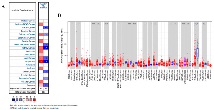
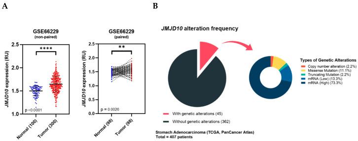
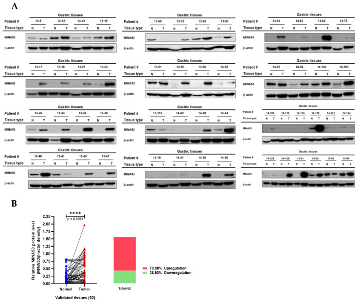
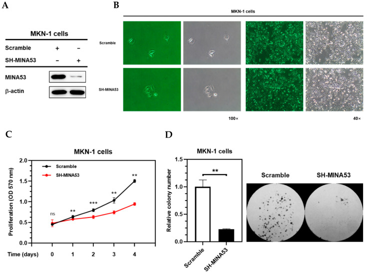
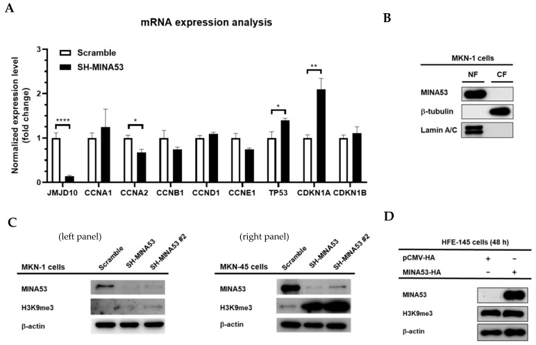
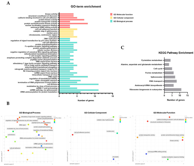
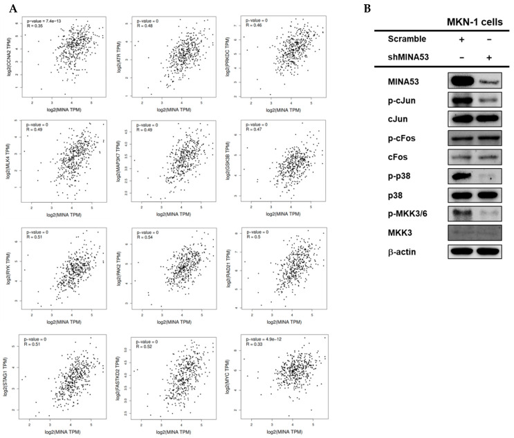
The JMJD10 gene and its encoded protein MYC-induced nuclear antigen (MINA53) are associated with multiple cancers. Besides having both an oncogenic and tumor suppressor function, the intricate role of JMJD10 in cancer is complex as it depends on the cancer type. In particular, the functional role of JMJD10/MINA53 in gastric cancer has been poorly understood. In this study, we have unraveled the molecular signatures and functional roles of JMJD10/MINA53 in gastric cancer by multiple approaches, i.e., multi-omics bioinformatics study, analysis of human gastric cancer tissues, and studies in vitro using knockdown or overexpression strategies in gastric cancer cell lines. The results indicated that the JMJD10 gene and MINA53 protein are commonly overexpressed in cancer patients. JMJD10/MINA53 is involved in the regulation of proliferation and survival of gastric cancer by controlling cell cycle gene expression. These processes are highly associated with MINA53 enzymatic activity in the regulation of H3K9me3 methylation status and controlling activation of AP-1 signaling pathways. This highlights the oncogenic role of JMJD10/MINA53 in gastric cancer and opens the opportunity to develop therapeutic targeting of JMJD10/MINA53 in gastric cancer.

Keywords: JMJD10/MINA53; KDM; gastric cancer; histone demethylase.


Conflict of interest statement

The authors declare no conflict of interest.

Figures

Figure 1
Figure 1
JMJD10 gene expression is upregulated in diverse types of cancer. (A) Expression of the JMJD10 gene in common cancers vs. normal tissues using the Oncomine database. The graph, generated by Oncomine, represents the number of statistically significant datasets (p < 0.01) overexpressed (red) or underexpressed (blue) in JMJD10 mRNA (cancer vs. corresponding normal tissue). The threshold was designed with the following parameters: p-value < 0.01, fold change = 1.5, and gene rank = Top 10%; the numbers in the boxes indicate the number of analyses that met the thresholds. (B) Expression of JMJD10 in cancer vs. normal tissues in various TCGA tumors using the DiffExp module in TIMER.
Figure 2
Figure 2
JMJD10 gene expression in gastric tumor microarray GSE66229 and genetic alteration profiling of JMJD10 in stomach adenocarcinoma TCGA PanCancer Atlas 2018. (A) Pairwise and non-pairwise comparison of JMJD10 expression between normal vs. tumor from dataset GSE66229 (RU: Relative Unit). (B) Alteration frequency analysis of the JMJ10 gene in stomach adenocarcinoma patients from TCGA PanCancer Atlas database extracted from the cBioPortal.
Figure 3
Figure 3
MINA53 protein expression analysis in human gastric cancer tissues. (A) Immunoblot results of human gastric cancer and adjacent normal tissues. (B) Pairwise analysis and box-plot overview of the relative MINA53 protein expression comparison from validated human gastric cancer tissue samples; normalized expression was done by measuring the relative density of MINA53/β-actin immunoblot results. Statistical significance was calculated using the paired t-test. The whole Western blot images please find in Figure S1.
Figure 4
Figure 4
The silencing of MINA53 inhibited proliferation and survival in gastric cancer cell lines. (A) Knockdown level of MINA-53 silenced MKN-1 cells. (B) Morphology of MINA53-silenced MKN-1 cells compared to scramble MKN-1 cells. (C) Proliferation assay of MINA53-silenced MKN-1 cells using MTT assay. (D) Colony formation of MINA53-silenced MKN-1 cells after 8 days. The whole Western blot images please find in Figure S1.
Figure 5
Figure 5
Molecular signature of MINA53 in gastric cancer cell lines. (A) mRNA expression analysis of JMJD10 in MINA53-silenced MKN-1 cells. (B) MINA53 mainly localized in the nuclear fraction; Lamin A/C and β-tubulin were used as loading controls for nuclear and cytosolic fractions, respectively. (C) Analysis of H3K9me3 level in MINA53-silenced MKN-1 cells (left panel) and MKN-45 cells (right panel). (D) Overexpression of MINA53 in human gastric normal cell lines (HFE-145) does not affect the H3K9me3 level. The whole Western blot images please find in Figure S1.
Figure 6
Figure 6
Functional enrichment analysis of JMJD10 in stomach adenocarcinoma. (A) Box plot representing enriched GO terms includes GO molecular function, cellular component, and biological process enrichment. (B) Semantic similarity-based scatterplots of GO-term enrichment results; the axes have no intrinsic meaning; semantically similar GO terms are closer together in the plot. (C) Box plot representing KEGG pathway enrichment results.
Figure 7
Figure 7
Regulatory role of MINA53 in AP-1 signaling in gastric cancer cell lines. (A) Gene expression correlation with JMJD10 in the STAD tumor dataset using GEPIA. (B) Immunoblot analysis of AP-1 signaling activation in MINA53-silenced MKN-1 cells. The whole Western blot images please find in Figure S1.

References

    1. Oh S., Shin S., Janknecht R. The small members of the JMJD protein family: Enzymatic jewels or jinxes? Biochim. Biophys. Acta (BBA) Bioenerg. 2019;1871:406–418. doi: 10.1016/j.bbcan.2019.04.002. - DOI - PMC - PubMed
    1. Franci G., Ciotta A., Altucci L. The Jumonji family: Past, present and future of histone demethylases in cancer. Biomol. Concepts. 2014;5:209–224. doi: 10.1515/bmc-2014-0010. - DOI - PubMed
    1. Johansson C., Tumber A., Che K., Cain P., Nowak R., Gileadi C., Oppermann U. The roles of Jumonji-type oxygenases in human disease. Epigenomics. 2014;6:89–120. doi: 10.2217/epi.13.79. - DOI - PMC - PubMed
    1. Cloos P.A., Christensen J., Agger K., Helin K. Erasing the methyl mark: Histone demethylases at the center of cellular differentiation and disease. Genome Res. 2008;22:1115–1140. doi: 10.1101/gad.1652908. - DOI - PMC - PubMed
    1. Bundred J.R., Hendrix E., Coleman M. The emerging roles of ribosomal histidyl hydroxylases in cell biology, physiology and disease. Cell. Mol. Life Sci. 2018;75:4093–4105. doi: 10.1007/s00018-018-2903-z. - DOI - PMC - PubMed

LinkOut - more resources