

XPS Modeling of Immobilized Recombinant Angiogenin and Apoliprotein A1 on Biodegradable Nanofibers
- PMID: 32370165
- PMCID: PMC7279301
- DOI: 10.3390/nano10050879
XPS Modeling of Immobilized Recombinant Angiogenin and Apoliprotein A1 on Biodegradable Nanofibers
Abstract
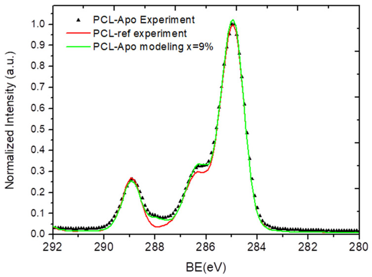
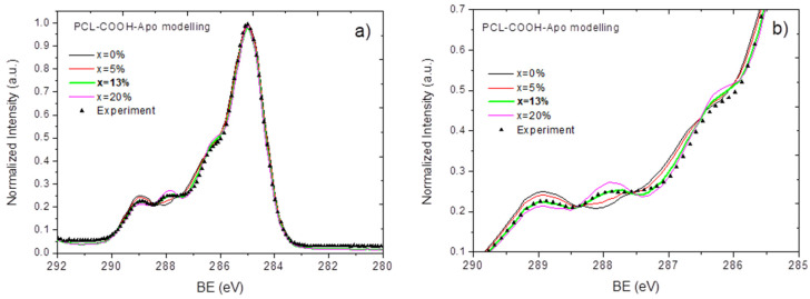
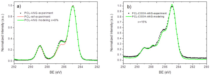
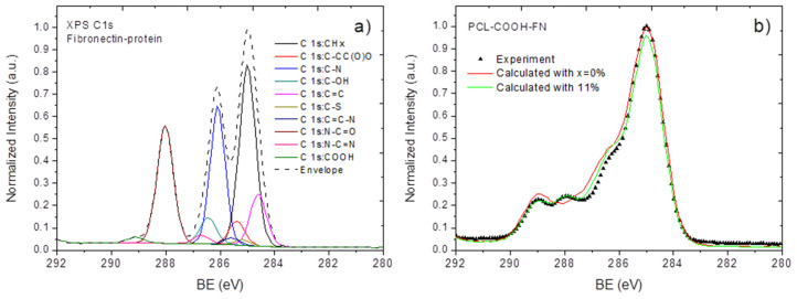
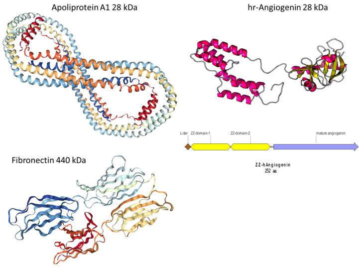
The immobilization of viable proteins is an important step in engineering efficient scaffolds for regenerative medicine. For example, angiogenin, a vascular growth factor, can be considered a neurotrophic factor, influencing the neurogenesis, viability, and migration of neurons. Angiogenin shows an exceptional combination of angiogenic, neurotrophic, neuroprotective, antibacterial, and antioxidant activities. Therefore, this protein is a promising molecule that can be immobilized on carriers used for tissue engineering, particularly for diseases that are complicated by neurotrophic and vascular disorders. Another highly important and viable protein is apoliprotein A1. Nevertheless, the immobilization of these proteins onto promising biodegradable nanofibers has not been tested before. In this work, we carefully studied the immobilization of human recombinant angiogenin and apoliprotein A1 onto plasma-coated nanofibers. We developed a new methodology for the quantification of the protein density of these proteins using X-ray photoelectron spectroscopy (XPS) and modeled the XPS data for angiogenin and apoliprotein A1 (Apo-A1). These findings were also confirmed by the analysis of immobilized Apo-A1 using fluorescent microscopy. The presented methodology was validated by the analysis of fibronectin on the surface of plasma-coated poly(ε-caprolactone) (PCL) nanofibers. This methodology can be expanded for other proteins and it should help to quantify the density of proteins on surfaces using routine XPS data treatment.
Keywords: X-ray photoelectron spectroscopy; angiogenin; biotechnology; nanofibers; plasma; polymers.
Conflict of interest statement
The authors declare no conflict of interest.
Figures
References
-
- Manakhov A.M., Landová M., Medalová J., Michlicek M., Polčak J., Nečas D., Zajíčková L. Cyclopropylamine plasma polymers for increased cell adhesion and growth. Plasma Process. Polym. 2016;14:1600123. doi: 10.1002/ppap.201600123. - DOI
-
- Solovieva A., Miroshnichenko S., Kovalskii A.M., Permyakova E.S., Popov Z., Dvořáková E., Kiryukhantsev-Korneev F.V., Obrosov A., Polčak J., Zajíčková L., et al. Immobilization of Platelet-Rich Plasma onto COOH Plasma-Coated PCL Nanofibers Boost Viability and Proliferation of Human Mesenchymal Stem Cells. Polymer. 2017;9:736. doi: 10.3390/polym9120736. - DOI - PMC - PubMed
-
- Zhang Z., Yoo R., Wells M., Beebe T.P., Biran R., Tresco P. Neurite outgrowth on well-characterized surfaces: Preparation and characterization of chemically and spatially controlled fibronectin and RGD substrates with good bioactivity. Biomaterials. 2005;26:47–61. doi: 10.1016/j.biomaterials.2004.02.004. - DOI - PubMed
Grants and funding
LinkOut - more resources
Full Text Sources
Miscellaneous
