. 2020 May 2;13(9):2108.
doi: 10.3390/ma13092108.

Eumelanin Precursor 2-Carboxy-5,6-Dihydroxyindole (DHICA) as Doping Factor in Ternary (PEDOT:PSS/Eumelanin) Thin Films for Conductivity Enhancement

Affiliations

Eumelanin Precursor 2-Carboxy-5,6-Dihydroxyindole (DHICA) as Doping Factor in Ternary (PEDOT:PSS/Eumelanin) Thin Films for Conductivity Enhancement

Ludovico Migliaccio et al. Materials (Basel). .

Abstract

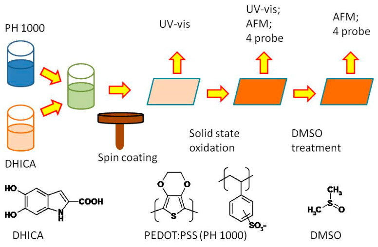
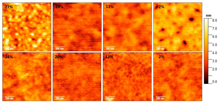
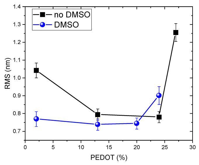
The integration of the pristine not-doped commercial poly(3,4-ethylenedioxythiophene):poly(styrenesulfonate) (PEDOT:PSS) PH1000 with eumelanin, the brown to black kind of melanin pigment, was achieved by dissolving the melanogenic precursors 2-carboxy-5,6-dihydroxyindole (DHICA) in the PH1000 suspension. Solid state oxidative polymerization of the catecholic indole allowed obtaining the ternary blend PEDOT:PSS/eumelanin. The introduction of DHICA into PH1000 produced a noticeable increase in the conductivity of PEDOT thin films akin to that produced by dimethyl sulfoxide (DMSO) treatment, opening up novel strategies for the simultaneous integration of eumelanin polymer and conductivity enhancement of PEDOT containing coatings, as well as the long term goal of replacing PSS by DHICA eumelanin for PEDOT pairing.

Keywords: PEDOT:PSS; bioinspired materials; conducting polymer; eumelanin; melanin.


Conflict of interest statement

The authors declare no conflict of interest. The funders had no role in the design of the study; in the collection, analyses, or interpretation of data; in the writing of the manuscript, or in the decision to publish the results.

Figures

Scheme 1
Scheme 1
Key steps of the carboxylated EuPH (C-EuPH) preparation and characterization. AFM, atomic force microscopy; DHICA, 2-carboxy-5,6-dihydroxyindole; PEDOT:PSS, poly(3,4-ethylenedioxythiophene):poly(styrenesulfonate).
Figure 1
Figure 1
Absorptivity in the UV/vis range of carboxylated EuPH (C-EuPH) samples. Thickness of all the films is in the range of 400–1000 nm (Table S1). It is evident how the increase in the 2-carboxy-5,6-dihydroxyindole (DHICA)-eumelanin content increases the absorption in the UV region of the films. PEDOT:PSS, poly(3,4-ethylenedioxythiophene):poly(styrenesulfonate).
Figure 2
Figure 2
The electrical conductivity of the films featuring various DHICA contents (DHICA weight fractions DHICA/(PEDOT + PSS + DHICA): 0, 0.055, 0.160, 0.195, 0.230, 0.265, 0.300, 0.405, 0.475, 0.545, 0.650, 0.755, 0.825). Data were measured before (triangle) and after (open square) DMSO treatment. The black full squares indicates the content of the PEDOT respect to all the components of the film, that is, proportional to PEDOT/(DHICA + PEDOT + PSS).
Figure 3
Figure 3
Conductivity increase in C-EuPH after DMSO treatment (left axis) and product of conductivity increase and the ratio of the conductivities before and after DMSO treatment (ΔCond × CondB/CondA).
Figure 4
Figure 4
Atomic force microscopy (AFM) topography of the investigated C-EuPH samples. Top panels refer to the samples not treated with DMSO and bottom panels refer to those prepared in DMSO. The PEDOT weight percent is reported in the top-left corner of each image, DHICA weight percent is as follows: PEDOT 27%–DHICA 6%; PEDOT 24%–DHICA 16%; PEDOT 20%–DHICA 30%; and PEDOT 2%–DHICA 94%, respectively. Scans are 1 µm2 with a resolution of 256 × 256 pixels.
Figure 5
Figure 5
Estimated root means square (RMS) of the samples surface for non-DMSO- and DMSO-treated samples. Lower percentages of the PEDOT are the result of higher contents of DHICA.

Similar articles

Cited by

References

    1. Malliaras G.G. Organic bioelectronics: A new era for organic electronics. Biochim. Biophys. Acta Gen. Subj. 2013;1830:4286–4287. doi: 10.1016/j.bbagen.2012.10.007. - DOI - PubMed
    1. Yuk H., Lu B., Zhao X. Hydrogel bioelectronics. Chem. Soc. Rev. 2019;48:1642–1667. doi: 10.1039/C8CS00595H. - DOI - PubMed
    1. Wang C., Huang Z., Xu S. Materials and Structures toward Soft Electronics. Adv. Mater. 2018;30:1801368. doi: 10.1002/adma.201801368. - DOI - PubMed
    1. Kumar A., Zhou C. The race to replace tin-doped indium oxide: Which material will win? Acs Nano. 2010;4:11–14. doi: 10.1021/nn901903b. - DOI - PubMed
    1. Lee H.B., Jin W.Y., Ovhal M.M., Kumar N., Kang J.W. Flexible transparent conducting electrodes based on metal meshes for organic optoelectronic device applications: A review. J. Mater. Chem. C. 2019;7:1087–1110. doi: 10.1039/C8TC04423F. - DOI

LinkOut - more resources