

Library of deep-learning image segmentation and outcomes model-implementations
- PMID: 32371142
- PMCID: PMC8474066
- DOI: 10.1016/j.ejmp.2020.04.011
Library of deep-learning image segmentation and outcomes model-implementations
Abstract
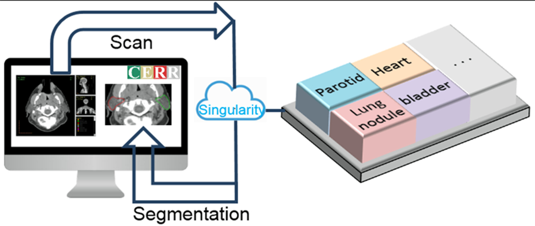
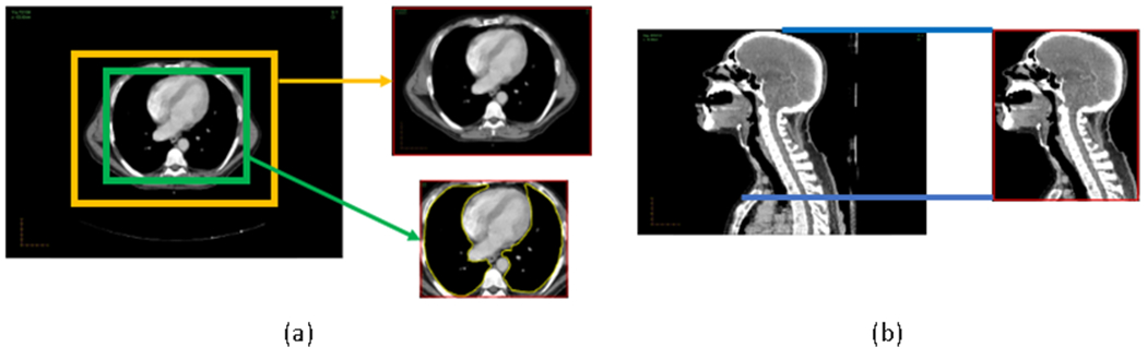
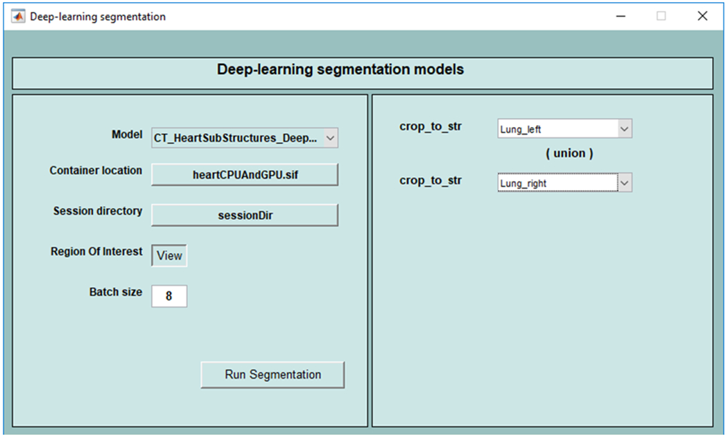
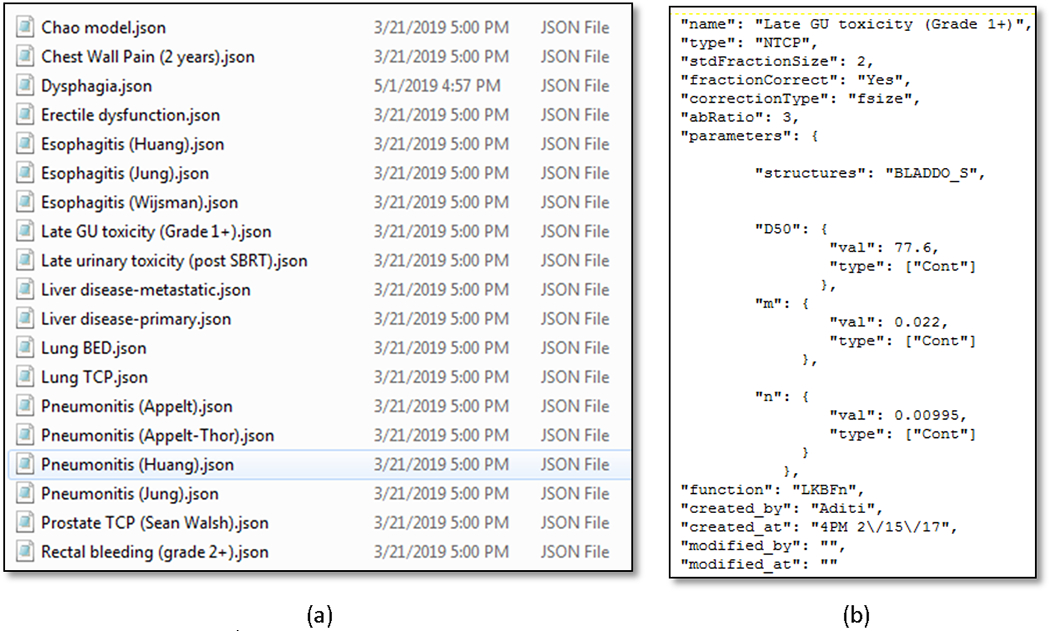
An open-source library of implementations for deep-learning-based image segmentation and outcomes models based on radiotherapy and radiomics is presented. As oncology treatment planning becomes increasingly driven by automation, such a library of model implementations is crucial to (i) validate existing models on datasets collected at different institutions, (ii) automate segmentation, (iii) create ensembles for improving performance and (iv) incorporate validated models in the clinical workflow. Inclusion of deep-learning-based image segmentation and outcomes models in the same library provides a fully automated and reproduceable pipeline to estimate prognosis. The library was developed with the Computational Environment for Radiological Research (CERR) software platform. Centralizing model implementations in CERR builds upon its rich set of radiotherapy and radiomics tools and caters to the world-wide user base. CERR provides well-validated feature extraction pipelines for radiotherapy dosimetry and radiomics with fine control over the calculation settings, allowing users to select appropriate parameters used in model derivation. Models for automatic image segmentation are distributed via containers, allowing them to be deployed with a variety of scientific computing architectures. The library includes implementations of popular DVH-based models outlined in the Quantitative Analysis of Normal Tissue Effects in the Clinic effort and recently published literature. Radiomics models include features from the Image Biomarker Standardization Initiative and application-specific features found to be relevant across multiple sites and image modalities. The library is distributed as a module within CERR at https://www.github.com/cerr/CERR under the GNU-GPL copyleft with additional restrictions on clinical and commercial use and provision to dual license in future.
Keywords: Deep-learning; Image segmentation; Library; Model implementations; Normal tissue complication; Radiomics; Radiotherapy outcomes; Tumor control.
Copyright © 2020. Published by Elsevier Ltd.
Figures
Similar articles
-
Technical Note: Extension of CERR for computational radiomics: A comprehensive MATLAB platform for reproducible radiomics research.Med Phys. 2018 Jun 13:10.1002/mp.13046. doi: 10.1002/mp.13046. Online ahead of print. Med Phys. 2018. PMID: 29896896 Free PMC article.
-
Deep learning for fully automated tumor segmentation and extraction of magnetic resonance radiomics features in cervical cancer.Eur Radiol. 2020 Mar;30(3):1297-1305. doi: 10.1007/s00330-019-06467-3. Epub 2019 Nov 11. Eur Radiol. 2020. PMID: 31712961
-
MIScnn: a framework for medical image segmentation with convolutional neural networks and deep learning.BMC Med Imaging. 2021 Jan 18;21(1):12. doi: 10.1186/s12880-020-00543-7. BMC Med Imaging. 2021. PMID: 33461500 Free PMC article.
-
Applications of radiomics and machine learning for radiotherapy of malignant brain tumors.Strahlenther Onkol. 2020 Oct;196(10):856-867. doi: 10.1007/s00066-020-01626-8. Epub 2020 May 11. Strahlenther Onkol. 2020. PMID: 32394100 Free PMC article. Review.
-
Machine and deep learning methods for radiomics.Med Phys. 2020 Jun;47(5):e185-e202. doi: 10.1002/mp.13678. Med Phys. 2020. PMID: 32418336 Free PMC article. Review.
Cited by
-
Treatment-integrated imaging, radiomics, and personalised radiotherapy: the future is at hand.Rep Pract Oncol Radiother. 2022 Sep 19;27(4):734-743. doi: 10.5603/RPOR.a2022.0071. eCollection 2022. Rep Pract Oncol Radiother. 2022. PMID: 36196410 Free PMC article. Review.
-
Longitudinal assessment of quality assurance measurements in a 1.5 T MR-linac: Part II-Magnetic resonance imaging.J Appl Clin Med Phys. 2022 Jun;23(6):e13586. doi: 10.1002/acm2.13586. Epub 2022 Mar 25. J Appl Clin Med Phys. 2022. PMID: 35332990 Free PMC article.
-
Interpretable Machine Learning for Choosing Radiation Dose-volume Constraints on Cardio-pulmonary Substructures Associated with Overall Survival in NRG Oncology RTOG 0617.Int J Radiat Oncol Biol Phys. 2023 Dec 1;117(5):1270-1286. doi: 10.1016/j.ijrobp.2023.06.009. Epub 2023 Jun 19. Int J Radiat Oncol Biol Phys. 2023. PMID: 37343707 Free PMC article. Clinical Trial.
-
Dermoscopic Image Classification of Pigmented Nevus under Deep Learning and the Correlation with Pathological Features.Comput Math Methods Med. 2022 May 28;2022:9726181. doi: 10.1155/2022/9726181. eCollection 2022. Comput Math Methods Med. 2022. PMID: 35669372 Free PMC article. Clinical Trial.
-
Using Auto-Segmentation to Reduce Contouring and Dose Inconsistency in Clinical Trials: The Simulated Impact on RTOG 0617.Int J Radiat Oncol Biol Phys. 2021 Apr 1;109(5):1619-1626. doi: 10.1016/j.ijrobp.2020.11.011. Epub 2020 Nov 13. Int J Radiat Oncol Biol Phys. 2021. PMID: 33197531 Free PMC article.
References
-
- LeCun Y, Bengio Y, Hinton G. Deep learning. Nature. 2015;521:436–44. - PubMed
-
- Van Zee KJ, Manasseh D-ME, Bevilacqua JLB, Boolbol SK, Fey JV, Tan LK, et al.A Nomogram for Predicting the Likelihood of Additional Nodal Metastases in Breast Cancer Patients With a Positive Sentinel Node Biopsy. Annals of Surgical Oncology. 2003;10:1140–51. - PubMed
MeSH terms
Grants and funding
LinkOut - more resources
Full Text Sources
Research Materials
