Review
. 2020 Oct;26(10):936-952.
doi: 10.1016/j.molmed.2020.03.012. Epub 2020 May 1.

Cell-to-Cell Transmission of Tau and α-Synuclein

Affiliations
Review

Cell-to-Cell Transmission of Tau and α-Synuclein

Norihito Uemura et al. Trends Mol Med. 2020 Oct.

Abstract

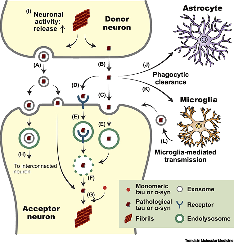
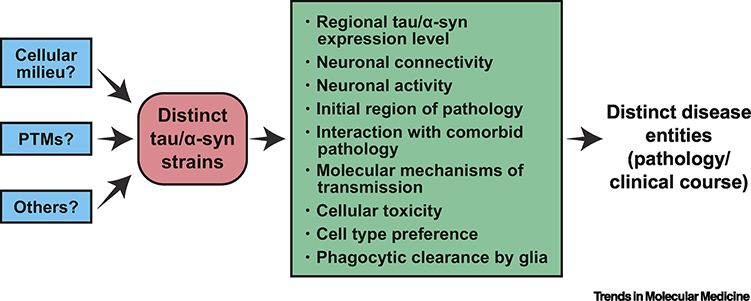
The stereotypical spread of pathological protein inclusions and clinicopathological heterogeneity are well described in neurodegenerative diseases. Accumulating evidence suggests that the former can be attributed to consecutive cell-to-cell transmission of pathological proteins between anatomically connected brain regions, while the latter has been hypothesized to result from the spread of conformationally distinct pathological protein aggregates, or strains. These emerging concepts have dramatically changed our understanding of neurodegenerative diseases. In this review, we first summarize the background and recent findings underpinning these concepts with a focus on two major pathological proteins: tau and α-synuclein. We then discuss their clinical implications for tauopathies and synucleinopathies and propose a working hypothesis for future research.

Keywords: Alzheimer’s disease; Parkinson’s disease; propagation; strains; synucleinopathies; tauopathies.


Figures

Figure 1.
Figure 1.. Distinct pathology in tauopathies and synucleinopathies
Phosphorylated tau immunohistochemistry (PHF1) (a–e,g,h). Hematoxylin-eosin staining (f). Misfolded α-syn immunohistochemistry (Syn303) (i–l). (a,b) NFTs and NP in the hippocampus of an AD subject. (c) Pick bodies in the dentate gyrus of a PiD subject. (d) Tufted astrocyte in the frontal cortex of a PSP subject. (e) Globose tangles in the medulla oblongata of a PSP subject. (f) Ballooned neuron in the amygdala of a CBD subject. (g,h) Coiled bodies and astrocytic plaque in the angular cortex of a CBD subject. (i) LB in a pigmented neuron in the SNpc of a PD subject. (j) LBs in the cingulate cortex of a DLB subject. (k) GCIs in the cerebellum of a MSA subject. (l) NI in the medulla oblongata of a MSA subject. Scale bar 40 μm.
Figure 2.
Figure 2.. Molecular mechanisms of cell-to-cell transmission
Schematic representation of cell-to-cell transmission of pathological tau and α-syn. Molecular mechanisms related to transmission of pathology are summarized in the context of neuron-to-neuron transmission. (a) Exosomal release of pathological tau [58, 59] and α-syn [60]. (b) Direct translocation of pathological tau [63]. (c) Macropinocytosis of pathological tau and α-syn [65]. (d) Receptor-mediated endocytosis of pathological α-syn [64, 66]. (e) Transportation of pathological tau [67] and α-syn [68] into endolysososmes. (f) Release of pathological tau and α-syn into cytosol through ruptured lysosomes [69]. (g) Templated misfolding of endogenous monomeric tau or α-syn. (h) Some exosomes containing pathological tau are incorporated in endosomes and are transmitted to the inter-connected neuron of the acceptor neuron [70]. (i) Enhanced neuronal activity promotes release pathological tau though the molecular mechanisms remain unclear [75, 77]. (j) Phagocytic clearance of pathological tau [79] and α-syn [80] by astrocytes. (k) Phagocytic clearance of pathological tau [–83] and α-syn [84] by microglia. (l) Microglia release exosomes containing pathological tau and promote transmission [61].
Figure 3.
Figure 3.. Formation and transmission of glial pathology
Schematic representation of the formation and transmission of glial pathology. Arrows indicate the direction of transmission of tau or α-syn pathology. (a) Neuron-to-astrocyte transmission of tau pathology [40, 85]. (b) Axon-to-oligodendroglia transmission of α-syn pathology [46]. (c) Oligodendroglia-to-oligodendroglia transmission of tau and α-syn pathology [46, 85].
Figure 4.
Figure 4.. Working hypothesis to investigate the pathological mechanisms of distinct tauopathies and synucleinopahies
Several factors highlighted in green define spread of tau and α-syn pathology in brains. These factors differently influence spread of pathology among distinct strains, thereby causing distinct disease entities. A few factors highlighted in blue could be the causes of distinct tau and α-syn strains, but the origin of strains remains largely unknown.
Figure I.
Figure I.
Structure and Intraneuronal Localization of Tau and α-Syn.
Figure II.
Figure II.
Aggregation and Molecular Entities of Tau and α-Syn.
Figure III.
Figure III.
Spreading Direction of Tau and α-Syn Pathology.

References

    1. Brettschneider J et al. (2015) Spreading of pathology in neurodegenerative diseases: a focus on human studies. Nat Rev Neurosci 16 (2), 109–20. - PMC - PubMed
    1. Hyman BT et al. (2012) National Institute on Aging-Alzheimer’s Association guidelines for the neuropathologic assessment of Alzheimer’s disease. Alzheimers Dement 8 (1), 1–13. - PMC - PubMed
    1. Braak H et al. (2006) Staging of Alzheimer disease-associated neurofibrillary pathology using paraffin sections and immunocytochemistry. Acta Neuropathol 112 (4), 389–404. - PMC - PubMed
    1. Thal DR et al. (2002) Phases of A beta-deposition in the human brain and its relevance for the development of AD. Neurology 58 (12), 1791–800. - PubMed
    1. Uchihara T et al. (2003) Pick body disease and Pick syndrome. Neuropathology 23 (4), 318–26. - PubMed

Publication types