. 2020 Jul 10;295(28):9299-9312.
doi: 10.1074/jbc.RA120.012987. Epub 2020 May 5.

Cooperative binding of the tandem WW domains of PLEKHA7 to PDZD11 promotes conformation-dependent interaction with tetraspanin 33

Affiliations

Cooperative binding of the tandem WW domains of PLEKHA7 to PDZD11 promotes conformation-dependent interaction with tetraspanin 33

Florian Rouaud et al. J Biol Chem. .

Abstract

Pleckstrin homology domain-containing A7 (PLEKHA7) is a cytoplasmic protein at adherens junctions that has been implicated in hypertension, glaucoma, and responses to Staphylococcus aureus α-toxin. Complex formation between PLEKHA7, PDZ domain-containing 11 (PDZD11), tetraspanin 33, and the α-toxin receptor ADAM metallopeptidase domain 10 (ADAM10) promotes junctional clustering of ADAM10 and α-toxin-mediated pore formation. However, how the N-terminal region of PDZD11 interacts with the N-terminal tandem WW domains of PLEKHA7 and how this interaction promotes tetraspanin 33 binding to the WW1 domain is unclear. Here, we used site-directed mutagenesis, glutathione S-transferase pulldown experiments, immunofluorescence, molecular modeling, and docking experiments to characterize the mechanisms driving these interactions. We found that Asp-30 of WW1 and His-75 of WW2 interact through a hydrogen bond and, together with Thr-35 of WW1, form a binding pocket that accommodates a polyproline stretch within the N-terminal PDZD11 region. By strengthening the interactions of the ternary complex, the WW2 domain stabilized the WW1 domain and cooperatively promoted the interaction with PDZD11. Modeling results indicated that, in turn, PDZD11 binding induces a conformational rearrangement, which strengthens the ternary complex, and contributes to enlarging a "hydrophobic hot spot" region on the WW1 domain. The last two lipophilic residues of tetraspanin 33, Trp-283 and Tyr-282, were required for its interaction with PLEKHA7. Docking of the tetraspanin 33 C terminus revealed that it fits into the hydrophobic hot spot region of the accessible surface of WW1. We conclude that communication between the two tandem WW domains of PLEKHA7 and the PLEKHA7-PDZD11 interaction modulate the ligand-binding properties of PLEKHA7.

Keywords: ADAM metallopeptidase domain 10 (ADAM10); PDZ domain containing 11 (PDZD11); PDZD11; PLEKHA7; Pleckstrin homology domain containing A7 (PLEKHA7); WW domain; adherens junction; cell-cell junction; cooperativity; membrane protein; molecular docking; polyproline; protein complex; tetraspanin 33; tetraspanin33; α-toxin.


Conflict of interest statement

Conflict of interest—The authors declare that they have no conflict of interest with the contents of this article.

Figures
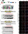
Figure 1.
Figure 1.
The WW2 domain of PLEKHA7 stabilizes the WW1 domain and rescues the interaction of WW1 mutants with PDZD11. A, schematic diagrams of PDZD11 (top) and PLEKHA7 (bottom), with the indicated structural domains. The N-terminal sequence of PDZD11 (P7-ID: PLEKHA7-interaction domain) interacts with the WW1 domain of PLEKHA7 (arrow) (15). B, top: sequence of the WW1 domain of PLEKHA7. Bottom: Weblogo diagram of residue conservation. C, top: sequence of WW1 domain, with mutations of highly conserved residues (to Ala) highlighted in yellow. The numbers above each residue indicate residue number in the sequence. Bottom: immunoblot analysis of GST pulldowns using GST (G) fused to WT and mutant WW1 domains as bait, and either PDZD11-HA or CFP-HA as preys. GST and GST fusion baits are indicated in red, and preys are indicated in green. Ponceau S-stained blots below immunoblots show baits. Red arrows indicate baits showing no or little proteolytic degradation. D, top: sequence of WW1 (continuous line, residues 1–53) and WW2 (dotted line, residues 43–98) domains, with highlighted WW1 mutations. The sequence linking the WW1 to the WW2 domain is in a dotted box. Bottom, immunoblot analysis of GST pulldowns using GST fused to WT and mutant WW1 + WW2 domains as bait, and either PDZD11-HA or CFP-HA as preys. Ponceau S-stained blots below the immunoblots show baits. E, immunofluorescence localization of endogenous PDZD11 in PLEKHA7-KO mCCD cells rescued either with GFP, or with either WT or WW1 point mutants of GFP-tagged full-length PLEKHA7. Merged images show nuclei in blue (DAPI). Arrows indicate junctional labeling. Arrowheads indicate decreased/undetected junctional labeling. Bar = 20 μm.
Figure 2.
Figure 2.
Binding of PDZD11 to the cleft between WW1 and WW2 domains requires either Asp-30 or Thr-35 of WW1 and His-75 of WW2. A, top: sequence of WW1 + WW2 domains, with mutations in residues within WW1 and WW2 highlighted in yellow. The numbers above and below each residue indicate residue number in the sequence. Bottom: immunoblot analysis of GST pulldowns using GST (G) fused to WT and mutant WW1 + WW2 domains as bait, and either PDZD11-HA or CFP-HA as preys. B, immunofluorescence localization of endogenous PDZD11 in PLEKHA7-KO mCCD cells rescued with either GFP, or with either WT or WW1 point mutants or WW2 point mutants, or WW1 + WW2 point mutants of GFP-tagged full-length PLEKHA7. Merged images show nuclei in blue (DAPI). Arrows indicate junctional labeling and asterisks indicate reduced/undetectable labeling. Bar = 20 μm. C, model of interaction between the WW1 domain (gray, with surface representation) and WW2 domain (orange). The dotted black line shows the hydrogen bond occurring between His-75 and Asp-30. D, electrostatic potential surface (EPS) of the WW1 and WW2 domains. The color ramp is set with a minimum value of −0.2 (red) and a maximum of 0.2 (blue). The WW2 domain presents in transparency an orange molecular surface for the EPS = 0 instead of the white used for WW1 to help in distinguish of the two domains. The probe radius to define the accessible surface area was set to 1.4 Å. The image shows the molecule from different space orientations turning along the y and x axes. In the upper part of the panel, a violet rectangle indicates the pocket that accommodate PDZD11. In the panel below the interacting surface between the two domains is shown from the below (+ 90°) and upper (−90°) views, respectively. The WW2 and WW1 domains are not displayed to allow the visualization of the contact surface indicated by the black circle. Images were obtained using the Molecular Surface tool from Maestro (Schrödinger release 2016-4) program.
Figure 3.
Figure 3.
Polyproline stretches and tyrosine 18 in the N-terminal region of PDZD11 are required for its interaction with PLEKHA7. A, top: schematic domain organization of PDZD11, and schemes of truncated constructs used in GST pulldown assays, and their interaction (Bind P7-His) with full-length PLEKHA7 (indicated by +, ±, −). Bottom: immunoblot analysis of GST pulldowns using either GST (G), or GST fused to either full-length PDZD11 or the truncated constructs (shown on top) as baits, and full-length PLEKHA7-His as prey. Ponceau S-stained blots below the immunoblots show baits. B, sequence of the P7-ID domain (15) of PDZD11, and Weblogo diagram of residue conservation within this region. C, immunoblot analysis of GST pulldowns using GST (G) fused to either WT or mutated P7-ID N-terminal domain of PDZD11 as bait and PLEKHA7-His as prey. D, immunoblot analysis of GST pulldowns using either GST, or GST fused to either WT or mutated full-length PDZD11 as bait, and PLEKHA7-His as prey. Ponceau S-stained blots below immunoblots show baits. E, immunofluorescence localization of endogenous PLEKHA7 in WT mCCD cells, transiently transfected with either GFP, or WT or mutants GFP-tagged full-length PDZD11. Merged images show nuclei in blue (DAPI). Arrows indicate junctional labeling, and asterisks indicate decreased/undetectable junctional labeling. Bar = 20 μm. F, binding mode of the PDZD11 dodecapeptide (PAYENPPAWIPP) in the pocket formed by WW1–WW2 domains. PDZD11 is shown in magenta as licorice style, whereas the WW1 and WW2 domains are represented, respectively, in gray and orange as a new cartoon and transparent surface style. The cyan dotted line indicates the cation-π interaction of Tyr-18 with Arg-21 in the WW1 domain. Hydrophobic contacts in the WW2 domain are represented with orange curved lines (near Phe-64), which indicate the pocket accommodating the tandem prolines 26-27. The image was generated with VMD software.
Figure 4.
Figure 4.
Binding of PDZ11 to the WW1 + WW2 domains induces an increase in the size of a hydrophobic hot spot region on WW1, which docks the tetraspanin 33 C-terminal peptide. A and B, lipophilic potential of the surface of the WW1–WW2 domains in the absence (A) or presence (B) of PDZD11. PDZD11 (16-27 peptide) is represented in magenta, with a wide frames surface. The image shows the molecule from different space orientations turning along the y (+90°) and x (−45°) axis. An orange double arrow indicates the hydrophobic hot spot region of the WW1 domain. In the presence of PDZD11 the surface of the lipophilic surface is increased, thus potentially enhancing the binding of hydrophobic ligands. The image was generated using the Molcad tool from SybylX-2.1.1. °C. Top, schemes of truncated constructs of Tspan33 used in GST pulldown assays. Bottom, immunoblot analysis of GST pulldowns using GST (G) fused to either C-terminal Tspan33 (G-T33-Cterm) or the truncated constructs (shown on left) as baits, and either PLEKHA7-WW(1 + 2)-Myc or GFP-Myc as preys. D, binding of the C-term dodecapeptide Tspan-33 (NQQHRADFWY) on the lipophilic surface of the WW1 domain. The WW1–WW2-PDZD11 complex is represented as surface with the WW1–WW2 domains as lipophilic potential and PDZD11 as magenta wide frames. The Tspan33 peptide is represented by gray capped sticks and a transparent lipophilic surface, where the Tyr and Trp residues constitute a highly hydrophobic part of the peptide. On the right, molecular docking shows how the Tspan33 peptide perfectly fits the lipophilic surface. E and F, single and double mutants of the WW1 domain fail to bind to Tspan33. Immunoblot analysis of GST pulldowns using either GST or GST (G) fused to Tspan33 C terminus (G-T33-Cterm) as baits, and either PLEKHA7-WW(1 + 2)-Myc WT and mutant or GFP-Myc as preys. G and H, PDZD11 promotes binding of the single but not double mutants of WW1 + WW2 to Tspan33. Immunoblot analysis of GST pulldowns using either GST or GST-Tspan33 baits (indicated in red), with either WT or single or double mutant WW1 + WW2 preys (green) Ponceau S-stained blots show the amounts of recombinant proteins used as bait. The third protein (either PDZD11-HA or CFP-HA, negative control) for tri-molecular pulldowns is shown in blue (normalization shown in H).

Similar articles

Cited by

References

    1. Meng W., Mushika Y., Ichii T., and Takeichi M. (2008) Anchorage of microtubule minus ends to adherens junctions regulates epithelial cell-cell contacts. Cell 135, 948–959 10.1016/j.cell.2008.09.040 - DOI - PubMed
    1. Pulimeno P., Bauer C., Stutz J., and Citi S. (2010) PLEKHA7 is an adherens junction protein with a tissue distribution and subcellular localization distinct from ZO-1 and E-cadherin. PLoS ONE 5, e12207 10.1371/journal.pone.0012207 - DOI - PMC - PubMed
    1. Pulimeno P., Paschoud S., and Citi S. (2011) A role for ZO-1 and PLEKHA7 in recruiting paracingulin to tight and adherens junctions of epithelial cells. J. Biol. Chem. 286, 16743–16750 10.1074/jbc.M111.230862 - DOI - PMC - PubMed
    1. Paschoud S., Jond L., Guerrera D., and Citi S. (2014) PLEKHA7 modulates epithelial tight junction barrier function. Tissue Barriers 2, e28755 10.4161/tisb.28755 - DOI - PMC - PubMed
    1. Levy D., Ehret G. B., Rice K., Verwoert G. C., Launer L. J., Dehghan A., Glazer N. L., Morrison A. C., Johnson A. D., Aspelund T., Aulchenko Y., Lumley T., Köttgen A., Vasan R. S., Rivadeneira F., et al. (2009) Genome-wide association study of blood pressure and hypertension. Nat. Genet. 41, 677–687 10.1038/ng.384 - DOI - PMC - PubMed

Publication types

LinkOut - more resources