. 2020 May 5;5(3):e00017-20.
doi: 10.1128/mSystems.00017-20.

Isolation and Characterization of the Novel Phage JD032 and Global Transcriptomic Response during JD032 Infection of Clostridioides difficile Ribotype 078

Affiliations

Isolation and Characterization of the Novel Phage JD032 and Global Transcriptomic Response during JD032 Infection of Clostridioides difficile Ribotype 078

Tinghua Li et al. mSystems. .

Abstract

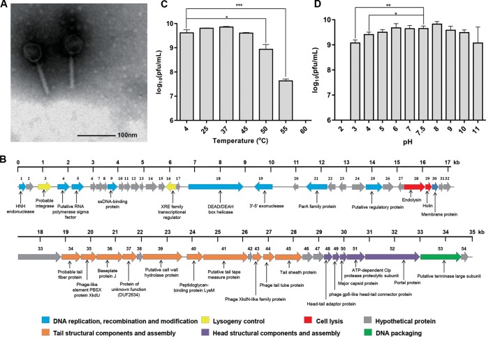
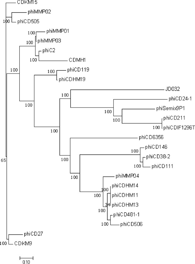
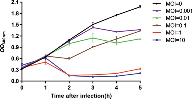
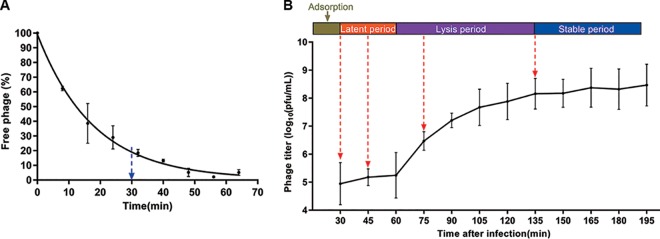
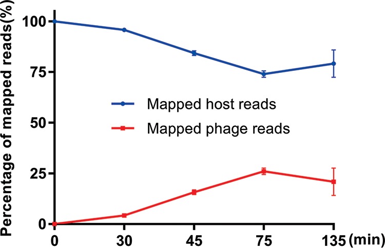
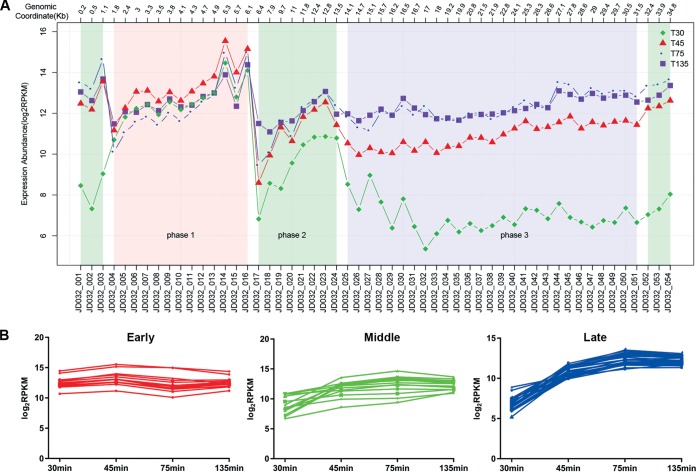
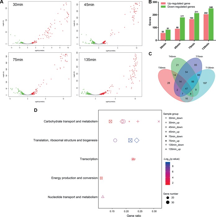
Insights into the interaction between phages and their bacterial hosts are crucial for the development of phage therapy. However, only one study has investigated global gene expression of Clostridioides (formerly Clostridium) difficile carrying prophage, and transcriptional reprogramming during lytic infection has not been studied. Here, we presented the isolation, propagation, and characterization of a newly discovered 35,109-bp phage, JD032, and investigated the global transcriptomes of both JD032 and C. difficile ribotype 078 (RT078) strain TW11 during JD032 infection. Transcriptome sequencing (RNA-seq) revealed the progressive replacement of bacterial host mRNA with phage transcripts. The expressed genes of JD032 were clustered into early, middle, and late temporal categories that were functionally similar. Specifically, a gene (JD032_orf016) involved in the lysis-lysogeny decision was identified as an early expression gene. Only 17.7% (668/3,781) of the host genes were differentially expressed, and more genes were downregulated than upregulated. The expression of genes involved in host macromolecular synthesis (DNA/RNA/proteins) was altered by JD032 at the level of transcription. In particular, the expression of the ropA operon was downregulated. Most noteworthy is that the gene expression of some antiphage systems, including CRISPR-Cas, restriction-modification, and toxin-antitoxin systems, was suppressed by JD032 during infection. In addition, bacterial sporulation, adhesion, and virulence factor genes were significantly downregulated. This study provides the first description of the interaction between anaerobic spore-forming bacteria and phages during lytic infection and highlights new aspects of C. difficile phage-host interactions.IMPORTANCE C. difficile is one of the most clinically significant intestinal pathogens. Although phages have been shown to effectively control C. difficile infection, the host responses to phage predation have not been fully studied. In this study, we reported the isolation and characterization of a new phage, JD032, and analyzed the global transcriptomic changes in the hypervirulent RT078 C. difficile strain, TW11, during phage JD032 infection. We found that bacterial host mRNA was progressively replaced with phage transcripts, three temporal categories of JD032 gene expression, the extensive interplay between phage-bacterium, antiphage-like responses of the host and phage evasion, and decreased expression of sporulation- and virulence-related genes of the host after phage infection. These findings confirmed the complexity of interactions between C. difficile and phages and suggest that phages undergoing a lytic cycle may also cause different phenotypes in hosts, similar to prophages, which may inspire phage therapy for the control of C. difficile.

Keywords: Clostridioides difficile; RNA-seq; bacteria-phage interaction; bacteriophage; ribotype 078; transcriptome.


Figures

FIG 1
FIG 1
Basic characteristics of phage JD032. (A) TEM of JD032. The diameter of the capsid and the length of the tail were 51.0 ± 1.70 nm and 90.0 ± 2.67 nm, respectively; these measurements were taken on 10 different particles. (B) Genome features of phage JD032. The predicted ORFs and their orientations are represented by arrows. The putative functional assignments are indicated below the ORFs. The functional modules were assigned based on gene annotation and genomic organization and are shown in different colors. (C) Thermostability of phage JD032. The x axis shows temperature, and the y axis shows the titer of phage JD032 after incubation for 1 h at different temperatures. (D) pH stability of phage JD032. The x axis shows pH values, and the y axis shows the titers of phage JD032 after incubation for 1 h at different pH values at 37°C. For panels C and D, data are displayed as the means plus standard deviations (SD) (error bars) from three independent experiments. *, 0.01 ≤ P < 0.05; **, 0.001 ≤ P < 0.01; and ***, P < 0.001, respectively.
FIG 2
FIG 2
Phylogenetic tree based on the whole genome of the C. difficile phage. The phylogenetic tree was generated using neighbor-joining analysis by MEGA-X. The genome of the C. difficile phage was downloaded from NCBI, and the accession numbers are shown in Table S2 in the supplemental material.
FIG 3
FIG 3
In vitro bactericidal activity of phage JD032 against C. difficile strain TW11. C. difficile strain TW11 was infected by phage JD032 at MOIs of 0.001, 0.01, 0.1, 1, and 10 and cultured for up to 5 h. Data are displayed as the means ± SD (error bars) from three independent experiments.
FIG 4
FIG 4
Lytic cycle of phage JD032 against C. difficile strain TW11. (A) Adsorption curve of JD032 to its host C. difficile TW11. The x axis shows the incubation time of JD032 and its host, and the y axis shows the percentage of the phage that did not adsorb to the host. (B) One-step growth curve of phage JD032. The x axis shows the incubation time of JD032 with its hosts after absorption for 30 min; the y axis shows the phage titers in the mixture at different times. Data are displayed as the means ± SD from three independent experiments.
FIG 5
FIG 5
Alignment of RNA read sets against the C. difficile (blue) or phage JD032 (red) genome at different time points after infection. Data are displayed as the means ± SD from three independent experiments.
FIG 6
FIG 6
Temporal kinetic transcriptional profile of phage JD032. (A) According to the expression abundance of phage genes at various time points, we divided these into three expression patterns. Genes that are highly expressed at 30 min (T30), 45 min (T45), and 75 min (T75) are called early, middle, and late genes, respectively. (B) Graphs displayed below the subclasses show expression profiles of the individual genes in that subclass as a function of time after infection.
FIG 7
FIG 7
Impact of phage JD032 infection on its host transcriptome. (A) Volcano plot of the C. difficile transcriptome following phage infection compared with the uninfected control. Each dot represents an open reading frame, with upregulated genes shown in red and downregulated genes in green. (B) Number and distribution of DEGs at different infection stages. (C) The Venn diagram shows the intersection of the number of DEGs at each time point. (D) Significant enrichment COG categories of host DEGs (up- and downregulated genes) at each time point after JD032 infection. The shape of the point indicates the time points. The enrichment q-value of each pathway was normalized as negative log10 P value and is shown as a color gradient. The number of genes enriched in each pathway is represented by the size of the points.
FIG 8
FIG 8
RT-qPCR verification of RNA-seq results. (A) Comparison of the expression levels of four phage genes measured by RT-qPCR and RNA-seq. RT-qPCR data were normalized using glutamate dehydrogenase (gluD) as an internal reference, and the relative expression level was calculated using the 2-ΔCт method. RNA-seq data were normalized to gene length and library size (RPKM). The replicates were averaged and presented as log10. (B andC) Verification using RT-qPCR for four DEGs (B), two TA system genes (C), one RM system gene (C), and one unaffected gene (C) of C. difficile strain TW11 upon infection. The relative expression levels of RT-qPCR data and RNA-seq were calculated using the 2-ΔΔCт method and fold change, respectively. The replicates were averaged and are presented as log10.

Similar articles

Cited by

References

    1. Bartlett JG, Gerding DN. 2008. Clinical recognition and diagnosis of Clostridium difficile infection. Clin Infect Dis 46(Suppl 1):S12–S18. doi:10.1086/521863. - DOI - PubMed
    1. Smits WK, Lyras D, Lacy DB, Wilcox MH, Kuijper EJ. 2016. Clostridium difficile infection. Nat Rev Dis Primers 2:16020. doi:10.1038/nrdp.2016.20. - DOI - PMC - PubMed
    1. Goorhuis A, Bakker D, Corver J, Debast SB, Harmanus C, Notermans DW, Bergwerff AA, Dekker FW, Kuijper EJ. 2008. Emergence of Clostridium difficile infection due to a new hypervirulent strain, polymerase chain reaction ribotype 078. Clin Infect Dis 47:1162–1170. doi:10.1086/592257. - DOI - PubMed
    1. Walker AS, Eyre DW, Wyllie DH, Dingle KE, Griffiths D, Shine B, Oakley S, O’Connor L, Finney J, Vaughan A, Crook DW, Wilcox MH, Peto TEA, Infections in Oxfordshire Research Database (IORD). 2013. Relationship between bacterial strain type, host biomarkers, and mortality in Clostridium difficile infection. Clin Infect Dis 56:1589–1600. doi:10.1093/cid/cit127. - DOI - PMC - PubMed
    1. Knetsch CW, Kumar N, Forster SC, Connor TR, Browne HP, Harmanus C, Sanders IM, Harris SR, Turner L, Morris T, Perry M, Miyajima F, Roberts P, Pirmohamed M, Songer JG, Weese JS, Indra A, Corver J, Rupnik M, Wren BW, Riley TV, Kuijper EJ, Lawley TD. 2017. Zoonotic transfer of Clostridium difficile harboring antimicrobial resistance between farm animals and humans. J Clin Microbiol 56:e01384-17. doi:10.1128/JCM.01384-17. - DOI - PMC - PubMed

LinkOut - more resources