. 2020 May 19;117(20):11126-11135.
doi: 10.1073/pnas.1920935117. Epub 2020 May 5.

Chronic mild hypoxia accelerates recovery from preexisting EAE by enhancing vascular integrity and apoptosis of infiltrated monocytes

Affiliations

Chronic mild hypoxia accelerates recovery from preexisting EAE by enhancing vascular integrity and apoptosis of infiltrated monocytes

Sebok K Halder et al. Proc Natl Acad Sci U S A. .

Abstract

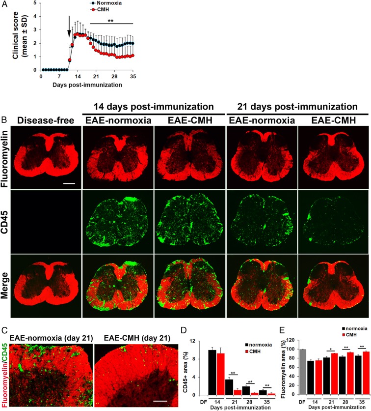
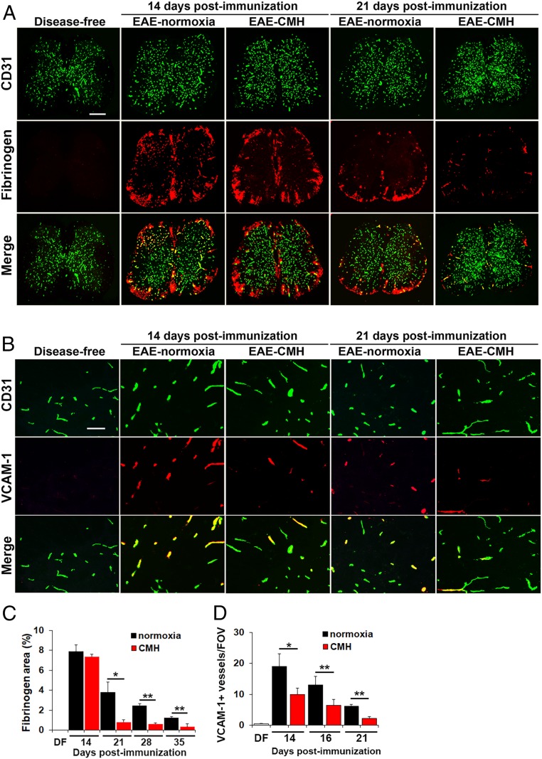
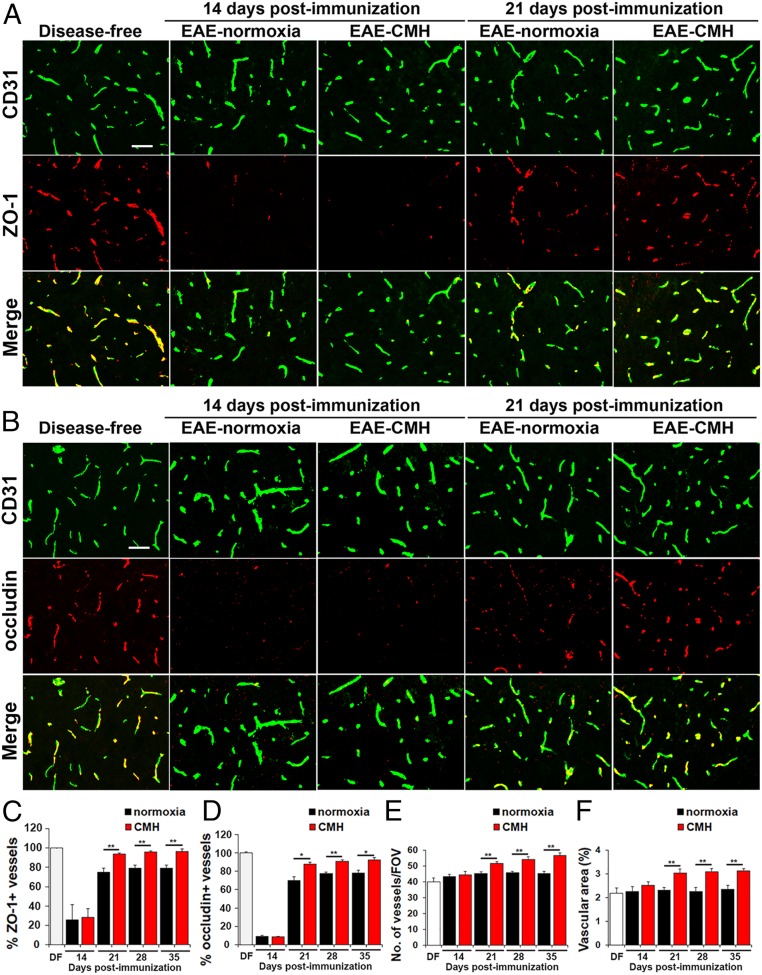
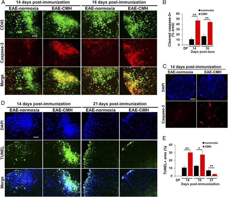
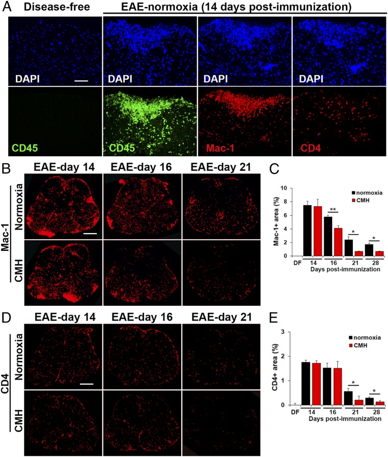
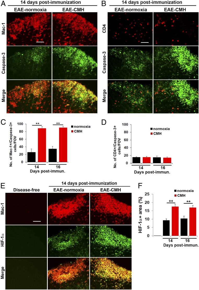
While several studies have shown that hypoxic preconditioning suppresses development of the experimental autoimmune encephalomyelitis (EAE) model of multiple sclerosis (MS), no one has yet examined the important clinically relevant question of whether mild hypoxia can impact the progression of preexisting disease. Using a relapsing-remitting model of EAE, here we demonstrate that when applied to preexisting disease, chronic mild hypoxia (CMH, 10% O2) markedly accelerates clinical recovery, leading to long-term stable reductions in clinical score. At the histological level, CMH led to significant reductions in vascular disruption, leukocyte accumulation, and demyelination. Spinal cord blood vessels of CMH-treated mice showed reduced expression of the endothelial activation molecule VCAM-1 but increased expression of the endothelial tight junction proteins ZO-1 and occludin, key mechanisms underlying vascular integrity. Interestingly, while equal numbers of inflammatory leukocytes were present in the spinal cord at peak disease (day 14 postimmunization; i.e., 3 d after CMH started), apoptotic removal of infiltrated leukocytes during the remission phase was markedly accelerated in CMH-treated mice, as determined by increased numbers of monocytes positive for TUNEL and cleaved caspase-3. The enhanced monocyte apoptosis in CMH-treated mice was paralleled by increased numbers of HIF-1α+ monocytes, suggesting that CMH enhances monocyte removal by amplifying the hypoxic stress manifest within monocytes in acute inflammatory lesions. These data demonstrate that mild hypoxia promotes recovery from preexisting inflammatory demyelinating disease and suggest that this protection is primarily the result of enhanced vascular integrity and accelerated apoptosis of infiltrated monocytes.

Keywords: blood–brain barrier; chronic mild hypoxia; experimental autoimmune encephalomyelitis; neuroinflammation; vascular integrity.


Conflict of interest statement

The authors declare no competing interest.

Figures

Fig. 1.
Fig. 1.
Chronic mild hypoxic treatment of preexisting EAE accelerates recovery at the clinical and histopathological levels. (A) The impact of CMH on clinical severity of relapsing–remitting EAE. Once mice developed a clinical score of 2 (arrow), they were randomly assigned to normoxic (control) or CMH conditions, and clinical score was evaluated at daily intervals. All points represent the mean ± SD (n = 26–32 mice per group, cumulative of three separate experiments). Note that compared to normoxic controls, mice treated with CMH showed accelerated clinical recovery, resulting in a marked and sustained reduction in long-term clinical score. (B and C) Frozen sections of lumbar spinal cord taken from disease-free, EAE–normoxia or EAE–CMH mice at the peak and remission phases of disease (14 and 21 d postimmunization, respectively) were stained for the inflammatory leukocyte marker CD45 (AlexaFluor-488) and fluoromyelin-red. (Scale bar, 500 μm [B] and 100 μm [C].) (D and E) Quantification of CD45 (D) and fluoromyelin (E) fluorescent signal at different timepoints. Results are expressed as the mean ± SEM percent area (n = 6 mice per group). Note that following peak disease, CMH markedly suppressed CD45+ leukocyte load within the spinal cord and protected against demyelination. *P < 0.05, **P < 0.01.
Fig. 2.
Fig. 2.
CMH treatment of preexisting EAE promotes beneficial changes in vascular integrity. (A and B) Frozen sections of lumbar spinal cord taken from disease-free, EAE–normoxia or EAE–CMH mice at the peak and remission phases of disease (14 and 21 d postimmunization, respectively) were stained for CD31 (AlexaFluor-488) and fibrinogen (Cy-3) in A (scale bar, 500 μm) or CD31 (AlexaFluor-488) and VCAM-1 (Cy-3) in B (scale bar, 50 μm). (C and D) Quantification of fibrinogen leakage, expressed as the mean ± SEM percent area (C) and VCAM-1 expression, expressed as the mean ± SEM number of VCAM-1+ vessels/FOV (D). n = 6 mice per group. Note that following peak disease, CMH markedly suppressed fibrinogen leakage and endothelial expression of VCAM-1. *P < 0.05, **P < 0.01.
Fig. 3.
Fig. 3.
CMH treatment of preexisting EAE promotes vascular remodeling and reexpression of endothelial tight junction proteins. (A and B) Frozen sections of lumbar spinal cord taken from disease-free, EAE–normoxia or EAE–CMH mice at the peak and remission phases of disease (14 and 21 d postimmunization, respectively) were stained for CD31 (AlexaFluor-488) and ZO-1 (Cy-3) in A or CD31 (AlexaFluor-488) and occludin (Cy-3) in B. (Scale bar, 50 μm.) (C and D) Quantification of ZO-1 (C) and occludin expression (D). Results are expressed as the mean ± SEM percent of vessels expressing ZO-1 or occludin. Note that during peak disease (14 d), both tight junction proteins were dramatically lost and were reexpressed during clinical remission (day 21), but at a faster rate in CMH-treated mice. (E and F) Quantification of changes in vessel density (E) and vascular area (F) during EAE progression under normoxic and CMH conditions. Results are expressed as the mean ± SEM of number of vessels/FOV or vascular area (percent of total). Note that CMH enhanced vessel density and vascular area at all timepoints from day 21 onward. n = 6 mice per group for all analyses. *P < 0.05, **P < 0.01.
Fig. 4.
Fig. 4.
CMH treatment of preexisting EAE promotes apoptosis of infiltrated leukocytes. (A, C, and D). Frozen sections of lumbar spinal cord taken from EAE–normoxia or EAE–CMH mice at the peak (14 and 16 d) and remission (21 d) phases of disease were stained for CD45 (AlexaFluor-488) and cleaved caspase-3 (Cy-3) in A, DAPI and cleaved caspase-3 (Cy-3) in C, or DAPI and TUNEL (AlexaFluor-488) in D. (Scale bar, 50 μm [A and D] and 500 μm [C].) (B and E) Quantification of cleaved caspase-3+ (B) and TUNEL+ (E) percent area. Results are expressed as the mean ± SEM (n = 6 mice per group). Note that during the peak phase of disease (days 14–16), CMH strongly enhanced the death of infiltrated leukocytes. *P < 0.05, **P < 0.01.
Fig. 5.
Fig. 5.
Monocytes are the predominant type of leukocyte within EAE lesions. (A, B, and D). Frozen sections of lumbar spinal cord taken from disease-free, EAE–normoxia or EAE–CMH mice at the peak (14 and 16 d) and remission (21 d) phases of disease were stained for DAPI, CD45 (AlexaFluor-488), Mac-1 (Cy-3), or CD4 (Cy-3) in A, Mac-1 (Cy-3) in B, or CD4 (Cy-3) in D. (Scale bar, 50 μm [A] or 500 μm [B and D].) (C and E) Quantification of the total Mac-1+ (C) or CD4+ (E) area. Results are expressed as the mean ± SEM percent area (n = 6 mice per group). Note that during the peak phase of disease (days 14–16), monocytes are the most abundant type of leukocyte in inflammatory lesions and that CMH strongly enhanced monocyte death during the remission phase. *P < 0.05, **P < 0.01.
Fig. 6.
Fig. 6.
CMH enhances apoptosis and HIF-1α expression in monocytes. (A, B, and E). Frozen sections of lumbar spinal cord taken from disease-free, EAE–normoxia or EAE–CMH mice at the peak phase of disease (14 d postimmunization) were stained for Mac-1 (Cy-3) and cleaved caspase-3 (AlexaFluor-488) in A, CD4 (Cy-3) and cleaved caspase-3 (AlexaFluor-488) in B, and Mac-1 (Cy-3) and HIF-1α (AlexaFluor-488) in E. (Scale bar, 25 μm.) (C, D and F). Quantification of the number of Mac-1/cleaved caspase-3 dual-positive (C) or CD4/cleaved caspase-3 dual-positive (D) cells/FOV or HIF-1α+ area (F). Results are expressed as the mean ± SEM (n = 6 mice per group). Note that during the peak phase of disease (days 14–16), CMH enhanced the death of infiltrated monocytes and increased HIF-1α expression in these cells. **P < 0.01.

References

    1. Doshi A., Chataway J., Multiple sclerosis, a treatable disease. Clin. Med. (Lond.) 17, 530–536 (2017). - PMC - PubMed
    1. Wingerchuk D. M., Carter J. L., Multiple sclerosis: Current and emerging disease-modifying therapies and treatment strategies. Mayo Clin. Proc. 89, 225–240 (2014). - PubMed
    1. Ffrench-Constant C., Pathogenesis of multiple sclerosis. Lancet 343, 271–275 (1994). - PubMed
    1. Lassmann H., “Multiple sclerosis pathology” in McAlpine’s Multiple Sclerosis, Compston A., Ed. (Churchill Livingstone, ed. 3, 1998), pp. 323–358.
    1. Dowden J., Corbett D., Ischemic preconditioning in 18- to 20-month-old gerbils: Long-term survival with functional outcome measures. Stroke 30, 1240–1246 (1999). - PubMed

Publication types

LinkOut - more resources