. 2020 May 19;117(20):10856-10864.
doi: 10.1073/pnas.1921848117. Epub 2020 May 5.

Direct observation of helicase-topoisomerase coupling within reverse gyrase

Affiliations

Direct observation of helicase-topoisomerase coupling within reverse gyrase

Xi Yang et al. Proc Natl Acad Sci U S A. .

Abstract

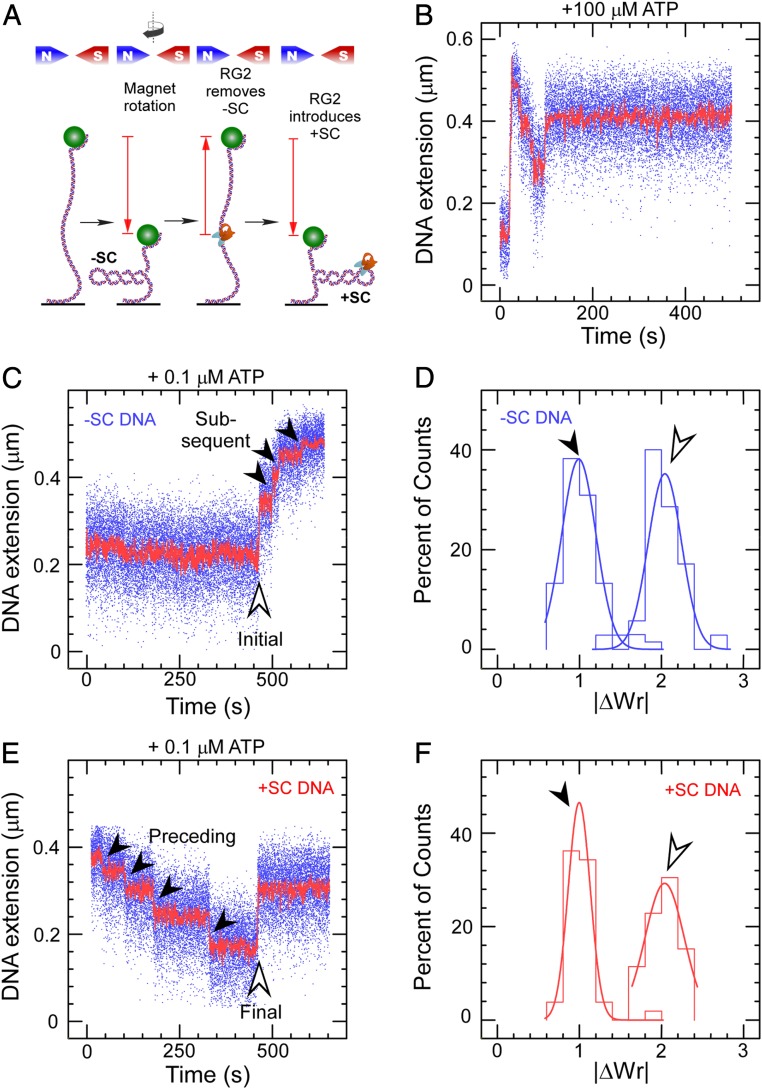
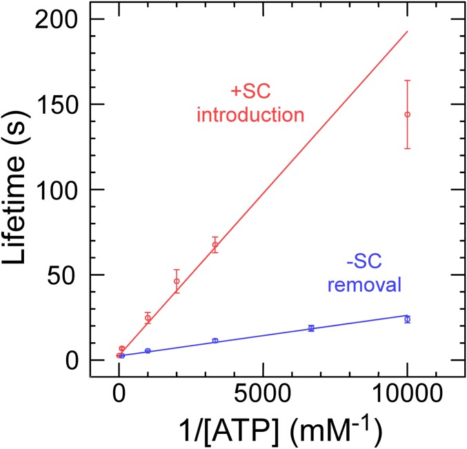
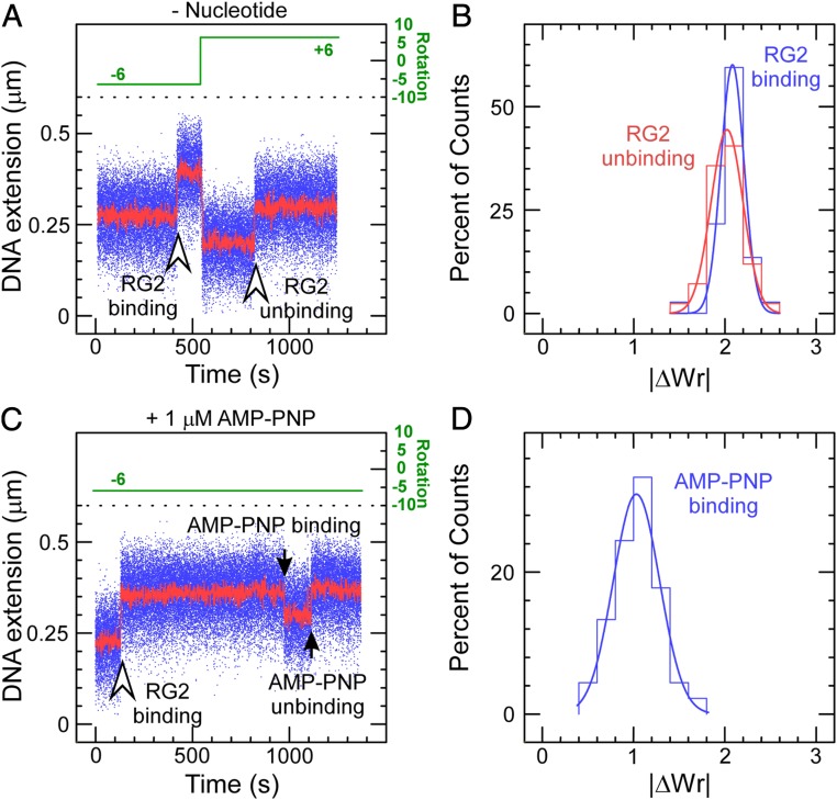
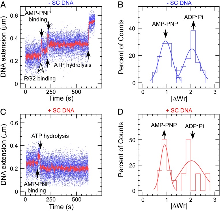
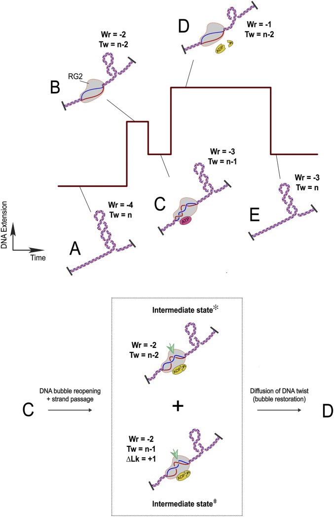
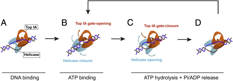
Reverse gyrases (RGs) are the only topoisomerases capable of generating positive supercoils in DNA. Members of the type IA family, they do so by generating a single-strand break in substrate DNA and then manipulating the two single strands to generate positive topology. Here, we use single-molecule experimentation to reveal the obligatory succession of steps that make up the catalytic cycle of RG. In the initial state, RG binds to DNA and unwinds ∼2 turns of the double helix in an ATP-independent fashion. Upon nucleotide binding, RG then rewinds ∼1 turn of DNA. Nucleotide hydrolysis and/or product release leads to an increase of 2 units of DNA writhe and resetting of the enzyme, for a net change of topology of +1 turn per cycle. Final dissociation of RG from DNA results in rewinding of the 2 turns of DNA that were initially disrupted. These results show how tight coupling of the helicase and topoisomerase activities allows for induction of positive supercoiling despite opposing torque.

Keywords: DNA topoisomerase; helicase; magnetic tweezers; single molecule.


Conflict of interest statement

The authors declare no competing interest.

Figures

Fig. 1.
Fig. 1.
Catalytic introduction of positive supercoils by RG2. (A) Sketch of the assay showing DNA tethered between a glass surface and a magnetic microsphere, which can be manipulated with a magnetic trap. Clockwise rotation of the magnets (as seen from above) results in negative DNA supercoils, which reduce DNA extension. RG2 first removes the negative supercoils before introducing net positive supercoiling. (B) Time trace for the extension of a negatively supercoiled DNA exposed to 50 pM RG2 and 100 µM ATP. (C) Time trace for the extension of a negatively supercoiled DNA exposed to RG2 and 0.1 µM ATP. The light arrow highlights the initial interaction, and the filled arrows, the subsequent interactions. (D) Histogram of change in DNA extension observed in C, taking into account both the initial and subsequent interaction observed between RG2 and DNA. Data are fit to Gaussian functions, respectively (solid line), with means ∆Wrinitial = 2.04 ± 0.04 (SEM; n = 35) and ΔWrsubsequent = 0.99 ± 0.03 (SEM; n = 68). E and F are as with the prior two panels but for positively supercoiled DNA, and with means ∆Wrpreceding = 1.00 ± 0.02 (SEM; n = 42) and ∆Wrfinal = −2.03 ± 0.07 (SEM; n = 21).
Fig. 2.
Fig. 2.
RG2 catalysis as a function of ATP concentration and sign of supercoiling. Lifetimes reflect average values ± SEM obtained from ∼65 to 269 individual events for removal of negative supercoils (blue) and introduction of net positive supercoils (red). Linear fits are to the Michaelis–Menten model (see text for details), returning for negative supercoiling 1/Vmax = 2.4 ± 0.2 s (SE) and KM = 1.0 ± 0.06 µM (SE) and for positive supercoiling 1/Vmax = 2.7 ± 0.2 s (SE) and KM = 7.1 ± 0.5 µM (SE).
Fig. 3.
Fig. 3.
RG2–DNA interactions in the absence and presence of AMP–PNP. (A) Time trace obtained in the absence of nucleotide cofactor shows RG2 binding to negatively supercoiled DNA results in an increase in DNA extension. This interaction can be reversed upon positive supercoiling, as evidenced by an increase in DNA extension observed in these conditions. (B) Histograms of change in DNA extension observed for negatively and positively supercoiled DNA, corresponding, respectively, to the DNA binding/unwinding step (blue) and DNA rewinding/dissociation step (red). The solid lines are Gaussian fits, indicating that unwinding involves a mean of 2.08 ± 0.02 turns of DNA (SEM; n = 37) and rewinding involves a mean of 2.02 ± 0.03 turns of DNA (SEM; n = 42). (C) Extension time trace obtained in the presence of 1 µM AMP–PNP shows RG2 binding to negatively supercoiled DNA and then binding nucleotide analog. This interaction spontaneously reverses. (D) Histogram of reversible extension changes observed in the presence of AMP–PNP. The solid line is a Gaussian fit giving a mean of 1.03 ± 0.04 (SEM; n = 45).
Fig. 4.
Fig. 4.
RG2–DNA interactions in the presence of a mixture of AMP–PNP and ATP. (A) Time trace obtained on negatively supercoiled DNA. Smaller steps with decrease in DNA extension correspond to AMP–PNP binding and the subsequent extension rebounds relate to ATP hydrolysis and Pi/ADP release. (B) Histogram of extension changes observed on negatively supercoiled DNA. Data are fit to a Gaussian for AMP–PNP binding and hydrolysis/product release, respectively, giving means ∆WrNTP = −0.95 ± 0.04 (SEM; n = 52) and ΔWrADP·Pi = 2.05 ± 0.07 (SEM; n = 34). (C) Time trace showing AMP–PNP binding and ATP hydrolysis/product release of RG2 obtained on positively supercoiled DNA, mirror symmetric with that on negative. The positively supercoiled DNA is obtained via rotating the magnets/magnetic bead immediately after having captured an RG2 binding to negatively supercoiled DNA. (D) Histogram of topological changes observed on positively supercoiled DNA. Data are fit to Gaussian functions (solid line), giving ∆WrNTP = −0.91 ± 0.08 (SEM; n = 10) and ΔWrADP·Pi = 2.03 ± 0.48 (SEM; n = 6).
Fig. 5.
Fig. 5.
Model for the RG cycle. RG2 is viewed as a topological state machine, manipulating DNA twist/writhe in an ordered reaction, through ATP-regulated helicase–topoisomerase coordination. (A) The starting point of an RG2 catalytic cycle is set with a DNA topological domain containing four negative DNA supercoils (Wr = −4; Tw = n). (B) RG2 binding unwinds 20 bp of DNA (Wr = −2; Tw = n − 2). (C) One-half of the 20-base DNA bubble is rewound due to ATP binding of RG2 (Wr = −3; Tw = n − 1). (Inset) In the subsequent ATP hydrolysis and product release stage (C to D), RG2 performs two steps: RG2 conducts a strand-passage reaction at the intersection point indicated by the green arrows (bottom half, Intermediate state; ΔLk = +1 → ΔWr = +1) and RG2 reopens the rewound DNA (top half, Intermediate state; ΔTw = −1 → ΔWr = +1). We do not know the order of the two intermediate steps and also the exact conformation of the base pairs. Together these transitions lead to a +2 unit change in DNA writhe. (D) RG2 finally reaches a nucleotide-free state after ATP hydrolysis and product release with the newly produced DNA twist diffused out of the enzyme and thus titrating out one negative supercoil on the DNA. (E) RG2 dissociation from DNA rewinds the 20-base DNA bubble, which returns two negative plectonemes back to the DNA substrate (Tw = −3; Wr = 0). Therefore, a complete ATP cycle by RG2 adds 1 unit of DNA linking number to the DNA (from AE).
Fig. 6.
Fig. 6.
Helicase–topoisomerase coupling in RG2 supercoiling. A second model of RG2’s catalytic cycle focuses on the RecQ–Top IA coupling. (A) RG2 unwinds a 20-bp DNA bubble. The open state of the helicase domain is coupled to a closed-gate conformation in the Top IA subunit. (B) ATP binding closes the helicase domain, causing rewinding of 10 bases of the DNA bubble, and opens the Top IA gate to rearrange ssDNA before strand passage. (C) ATP hydrolysis and/or subsequent product release reopens the helicase domain to regenerate the original 20-bp bubble, and this is coordinated with the DNA strand passage through the Top IA gate. (D) The RG2–DNA complex reset to the initial 20-bp DNA unwinding bubble can bind a new ATP and repeat its cycle.

Similar articles

Cited by

References

    1. Kikuchi A., Asai K., Reverse gyrase—a topoisomerase which introduces positive superhelical turns into DNA. Nature 309, 677–681 (1984). - PubMed
    1. Forterre P., Mirambeau G., Jaxel C., Nadal M., Duguet M., High positive supercoiling in vitro catalyzed by an ATP and polyethylene glycol-stimulated topoisomerase from Sulfolobus acidocaldarius. EMBO J. 4, 2123–2128 (1985). - PMC - PubMed
    1. Nakasu S., Kikuchi A., Reverse gyrase; ATP-dependent type I topoisomerase from Sulfolobus. EMBO J. 4, 2705–2710 (1985). - PMC - PubMed
    1. Nadal M., Mirambeau G., Forterre P., Reiter W. D., Duguet M., Positively supercoiled DNA in a virus-like particle of an archaebacterium. Nature 321, 256–258 (1986).
    1. Bell S. D., Jaxel C., Nadal M., Kosa P. F., Jackson S. P., Temperature, template topology, and factor requirements of archaeal transcription. Proc. Natl. Acad. Sci. U.S.A. 95, 15218–15222 (1998). - PMC - PubMed

Publication types

Supplementary concepts

LinkOut - more resources