. 2020 May 19;117(20):10806-10817.
doi: 10.1073/pnas.1920097117. Epub 2020 May 5.

Structural basis for divergent and convergent evolution of catalytic machineries in plant aromatic amino acid decarboxylase proteins

Affiliations

Structural basis for divergent and convergent evolution of catalytic machineries in plant aromatic amino acid decarboxylase proteins

Michael P Torrens-Spence et al. Proc Natl Acad Sci U S A. .

Abstract

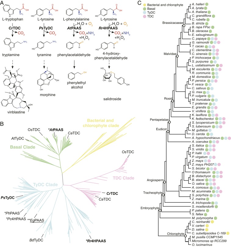
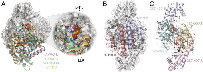
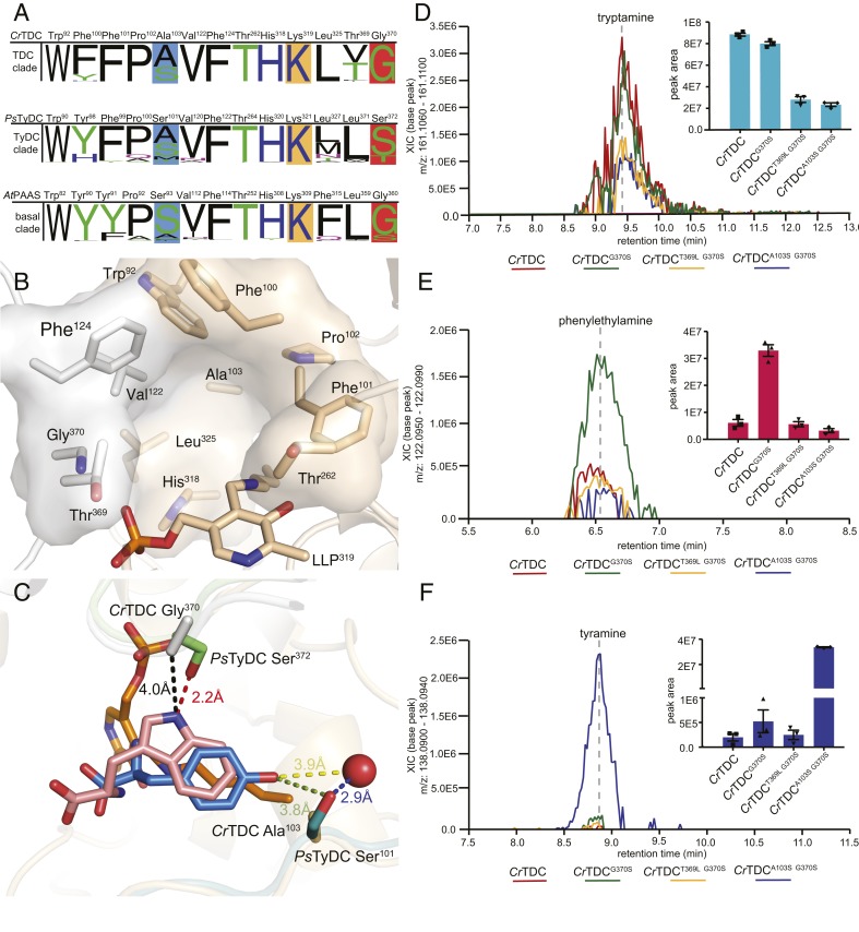
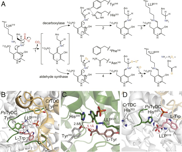
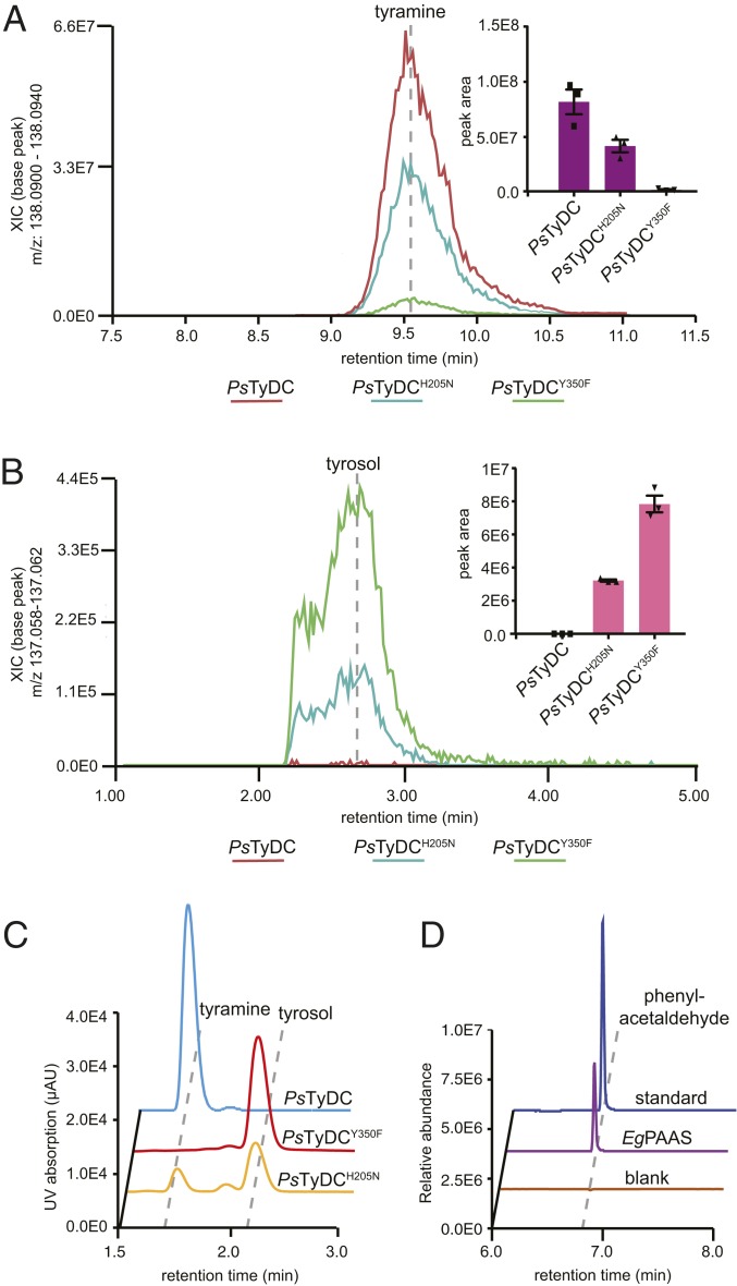
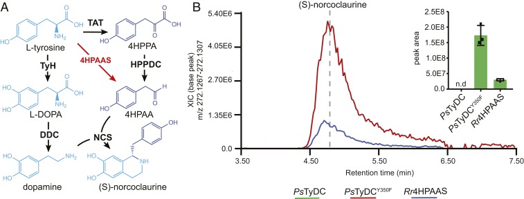
Radiation of the plant pyridoxal 5'-phosphate (PLP)-dependent aromatic l-amino acid decarboxylase (AAAD) family has yielded an array of paralogous enzymes exhibiting divergent substrate preferences and catalytic mechanisms. Plant AAADs catalyze either the decarboxylation or decarboxylation-dependent oxidative deamination of aromatic l-amino acids to produce aromatic monoamines or aromatic acetaldehydes, respectively. These compounds serve as key precursors for the biosynthesis of several important classes of plant natural products, including indole alkaloids, benzylisoquinoline alkaloids, hydroxycinnamic acid amides, phenylacetaldehyde-derived floral volatiles, and tyrosol derivatives. Here, we present the crystal structures of four functionally distinct plant AAAD paralogs. Through structural and functional analyses, we identify variable structural features of the substrate-binding pocket that underlie the divergent evolution of substrate selectivity toward indole, phenyl, or hydroxyphenyl amino acids in plant AAADs. Moreover, we describe two mechanistic classes of independently arising mutations in AAAD paralogs leading to the convergent evolution of the derived aldehyde synthase activity. Applying knowledge learned from this study, we successfully engineered a shortened benzylisoquinoline alkaloid pathway to produce (S)-norcoclaurine in yeast. This work highlights the pliability of the AAAD fold that allows change of substrate selectivity and access to alternative catalytic mechanisms with only a few mutations.

Keywords: AAAD; aromatic amino acid metabolism; enzyme evolution; specialized metabolism.


Conflict of interest statement

Competing interest statement: J.-K.W. is a cofounder, a member of the Scientific Advisory Board, and a shareholder of DoubleRainbow Biosciences, which develops biotechnologies related to natural products.

Figures

Fig. 1.
Fig. 1.
Function, phylogeny, and taxonomic distribution of plant AAADs. (A) Biochemical functions of four representative plant AAADs in the context of their native specialized metabolic pathways. The dashed arrows indicate multiple catalytic steps. (B) A simplified maximum likelihood phylogenetic tree of bacteria, chlorophyte, and plant AAADs. A fully annotated tree is shown in SI Appendix, Fig. S3. The bacterial/chlorophyte, basal, TyDC, and TDC clades are colored in yellow, green, blue, and pink, respectively. Functionally characterized enzymes are labeled at the tree branches. The four AAADs for which their crystal structures were resolved in this study are denoted in bold. The EgPAAS identified and characterized in this study is underlined; * and ** denote two mechanistic classes of AASs that harbor naturally occurring substitutions at the large-loop catalytic tyrosine or the small-loop catalytic histidine, respectively. (C) Taxonomic distribution of plant AAADs across major lineages of green plants. The tree illustrates the phylogenetic relationship among Phytozome V12 species with sequenced genomes. The presence of yellow, green, blue, or pink circles at the tree branches indicates the presence of one or more AAAD sequences from the bacterial/chlorophyte, basal, TyDC, or TDC clades, respectively.
Fig. 2.
Fig. 2.
The overall structure of plant AAADs. (A) An overlay of the CrTDC (orange), PsTyDC (green), AtPAAS (pink), and Rr4HPAAS (cyan) structures. All four structures exist as highly similar homodimers, but, for visual simplicity, the cartoon structures were only displayed for the bottom monomers. The top monomer of CrTDC is displayed in gray cartoon and surface representation. The dashed circle highlights the CrTDC active site which contains the l-tryptophan substrate and the prosthetic group LLP. (B) The CrTDC N-terminal segments from the two monomers, one colored in salmon and one in blue, form the hydrophobic dimer interface. The remainder of the homodimer is displayed in gray. (C) The configuration of the CrTDC middle (colored in teal and brown) and C-terminal segments (colored in blue and pink) from the two monomers. The N-terminal segments are displayed in transparent gray cartoons, and the prosthetic LLPs are circled and displayed as spheres. The models exhibited in B and C are in the same orientation, which is rotated 90° around the vertical axis from the view in A.
Fig. 3.
Fig. 3.
Active site pocket composition and residues that dictate substrate selectivity in plant AAADs. (A) Active-site-lining residues from plant AAADs were identified and queried for conservation against all AAAD homologs identified from the 93 Phytozome V12.1 annotated green plant genomes. The height of the residue label displays the relative amino acid frequency, excluding sequence gaps, in the basal, TyDC, and TDC clades. The position of the active site pocket residues from each clade are referenced against the AtPAAS, PsTyDC, and CrTDC sequences. Polar amino acids are colored in green, basic amino acids are colored in blue, acidic amino acids are colored in red, and hydrophobic amino acids are colored in black. Residues highlighted in blue and dark orange boxes denote residues involved in hydroxylated vs. unhydroxylated and phenolic vs. indolic substrate recognition, respectively. The conserved lysine residues, represented as the LLP prosthetic group in several crystal structures, are marked by a light orange box. (B) The CrTDC active-site pocket is composed of residues from both chain A and chain B, colored in beige and white, respectively. The pocket is composed of conserved nonpolar residues (Pro102, Val122, and Leu325), aromatic residues (Trp92, Phe100, Phe101, Phe124, and His318), and a polar residue (Thr262). Additionally, the active site contains three variable residues (Ala103, Thr369, and Gly370), which differ across different AAAD clades. (C) Superimposition of the substrate-complexed CrTDC and PsTyDC structures. CrTDC chain A and chain B are displayed in beige and white, respectively, while relevant portions of the PsTyDC chain A and chain B are displayed in green and deep teal, respectively. The l-tryptophan ligand from the CrTDC structure is colored in pink, while the PsTyDC l-tyrosine ligand is colored in blue. The red sphere represents a PsTyDC active-site water likely involved in substrate recognition. (DF) Relative in vivo decarboxylation of (D) ʟ-tryptophan, (E) l-phenylalanine, and (F) ʟ-tyrosine catalyzed by wild-type and various mutants of CrTDC as measured in transgenic yeast. The error bars indicate SEM based on biological triplicates while the squares, triangles, and diamonds represent the individual data points.
Fig. 4.
Fig. 4.
Catalytic mechanisms and conformational changes of plant AAAD proteins. (A) The proposed alternative PLP-mediated catalytic mechanisms for the canonical decarboxylase and derived aldehyde synthase in plant AAAD proteins. After transaldimation of the CrTDC internal aldimine to release the active-site Lys319, the PLP amino acid external aldimine loses the α-carboxyl group as CO2 to generate a quinonoid intermediate stabilized by the delocalization of the paired electrons (1). In a canonical decarboxylase (e.g., CrTDC), the carbanion at Cα is protonated by the acidic p-hydroxyl of Tyr348-B located on the large loop, which is facilitated by its neighboring His203-A located on the small loop (2). The CrTDC LLP319 internal aldimine is regenerated, accompanied by the release of the monoamine product (3). In an evolutionarily new aldehyde synthase, the Cα protonation step essential for the canonical decarboxylase activity is impaired when the large-loop catalytic tyrosine is mutated to phenylalanine (* as in AtPAAS), or when the small-loop catalytic histidine is mutated to asparagine (** as in EgPAAS), allowing the Cα carbanion to attack a molecular oxygen to produce a peroxide intermediate (4). This peroxide intermediate decomposes to give aromatic acetaldehyde, ammonia, and hydrogen peroxide products and regenerate the LLP internal aldimine (^ as in AtPAAS) (5). Aro represents the aromatic moiety of an aromatic l-amino acid. (B) An overlay of the PsTyDC structure with its large loop in a closed conformation (green) upon the CrTDC structure with its large loop in an open conformation (beige). (C) The closed conformation of PsTyDC active site displaying the catalytic machinery in a configuration ready to engage catalysis. Chain A is colored in white, chain B is colored in green, and the l-tyrosine substrate is displayed in dark pink. (D) Open and closed small-loop conformations observed in the CrTDC and PsTyDC structures. The PsTyDC small loop with the catalytic histidine is in a closed conformation (green), while the CrTDC structure exhibits its small-loop histidine in an open conformation (white).
Fig. 5.
Fig. 5.
Two alternative molecular strategies to arrive at aldehyde synthase chemistry from a canonical AAAD progenitor. Transgenic yeast strains expressing PsTyDCH205N and PsTyDCY350F display (A) reduced tyramine production but (B) elevated levels of tyrosol (reduced from 4HPAA by yeast endogenous metabolism) in comparison to transgenic yeast expressing wild-type PsTyDC. Cultures were grown and metabolically profiled in triplicate. The error bars in the bar graphs (Insets) indicate SEM and the squares and triangles represent the individual data points. (C) LC-UV chromatograms showing the relative decarboxylation and aldehyde synthase products produced by purified recombinant PsTyDC, PsTyDCY350F, or PsTyDCH205N enzymes when incubated with 0.5 mM l-tyrosine for 5, 25, and 50 min, respectively. After enzymatic reaction, the 4HPAA aldehyde product was chemically reduced by sodium borohydride to tyrosol prior to detection. (D) Phenylacetaldehyde formation from the incubation of purified recombinant EgPAAS with l-phenylalanine measured by GC-MS.
Fig. 6.
Fig. 6.
The utility of 4HPAAS in metabolic engineering of a shortened BIA pathway for (S)-norcoclaurine production in yeast. (A) Canonically proposed (S)-norcoclaurine biosynthetic pathway (black arrows) rerouted by the use of a 4HPAAS (red arrows). (B) Engineering of (S)-norcoclaurine production in yeast using two AAAD proteins with the 4HPAAS activity. All multigene vectors used to transform yeast contain BvTyH, PpDDC, and PsNCS in addition to either PsTyDC, PsTyDCY350F, or Rr4HPAAS. Cultures were grown and measured in triplicate. The error bars in the bar graph indicate SEM whereas the squares and triangles represent the individual data points; n.d., not detected.

Similar articles

Cited by

References

    1. Weng J.-K., Philippe R. N., Noel J. P., The rise of chemodiversity in plants. Science 336, 1667–1670 (2012). - PubMed
    1. Chae L., Kim T., Nilo-Poyanco R., Rhee S. Y., Genomic signatures of specialized metabolism in plants. Science 344, 510–513 (2014). - PubMed
    1. Kries H. et al. ., Structural determinants of reductive terpene cyclization in iridoid biosynthesis. Nat. Chem. Biol. 12, 6–8 (2016). - PMC - PubMed
    1. Kaltenbach M. et al. ., Evolution of chalcone isomerase from a noncatalytic ancestor. Nat. Chem. Biol. 14, 548–555 (2018). - PubMed
    1. Burkhard P., Dominici P., Borri-Voltattorni C., Jansonius J. N., Malashkevich V. N., Structural insight into Parkinson’s disease treatment from drug-inhibited DOPA decarboxylase. Nat. Struct. Biol. 8, 963–967 (2001). - PubMed

Publication types

MeSH terms

Substances