

Ultrastructural Evidence for Direct Renal Infection with SARS-CoV-2
- PMID: 32371536
- PMCID: PMC7460898
- DOI: 10.1681/ASN.2020040432
Ultrastructural Evidence for Direct Renal Infection with SARS-CoV-2
Erratum in
-
Correction.J Am Soc Nephrol. 2020 Oct;31(10):2494. doi: 10.1681/ASN.2020081117. J Am Soc Nephrol. 2020. PMID: 32999040 Free PMC article. No abstract available.
Abstract
Background: A significant fraction of patients with coronavirus disease 2019 (COVID-19) display abnormalities in renal function. Retrospective studies of patients hospitalized with COVID-19 in Wuhan, China, report an incidence of 3%-7% progressing to ARF, a marker of poor prognosis. The cause of the renal failure in COVID-19 is unknown, but one hypothesized mechanism is direct renal infection by the causative virus, SARS-CoV-2.
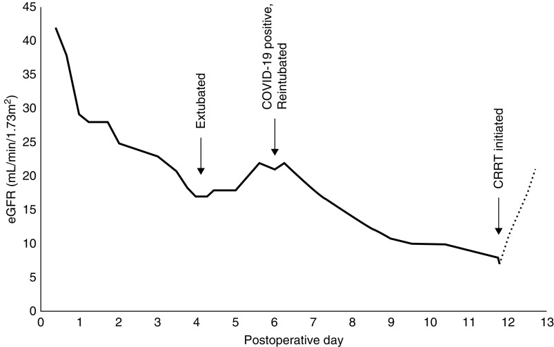
Methods: We performed an autopsy on a single patient who died of COVID-19 after open repair of an aortic dissection, complicated by hypoxic respiratory failure and oliguric renal failure. We used light and electron microscopy to examine renal tissue for evidence of SARS-CoV-2 within renal cells.
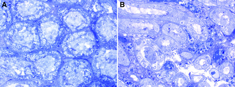
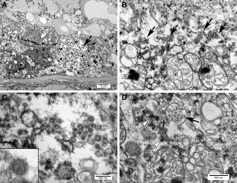
Results: Light microscopy of proximal tubules showed geographic isometric vacuolization, corresponding to a focus of tubules with abundant intracellular viral arrays. Individual viruses averaged 76 µm in diameter and had an envelope studded with crown-like, electron-dense spikes. Vacuoles contained double-membrane vesicles suggestive of partially assembled virus.
Conclusions: The presence of viral particles in the renal tubular epithelium that were morphologically identical to SARS-CoV-2, and with viral arrays and other features of virus assembly, provide evidence of a productive direct infection of the kidney by SARS-CoV-2. This finding offers confirmatory evidence that direct renal infection occurs in the setting of AKI in COVID-19. However, the frequency and clinical significance of direct infection in COVID-19 is unclear. Tubular isometric vacuolization observed with light microscopy, which correlates with double-membrane vesicles containing vacuoles observed with electronic microscopy, may be a useful histologic marker for active SARS-CoV-2 infection in kidney biopsy or autopsy specimens.
Keywords: COVID-19; SARS-CoV-2; acute kidney failure; autopsy; electron microscopy; renal pathology.
Copyright © 2020 by the American Society of Nephrology.
Figures
Comment in
-
Kidney Involvement in COVID-19: Need for Better Definitions.J Am Soc Nephrol. 2020 Sep;31(9):2224-2225. doi: 10.1681/ASN.2020050630. Epub 2020 Jul 9. J Am Soc Nephrol. 2020. PMID: 32646857 Free PMC article. No abstract available.
-
Caution in Identifying Coronaviruses by Electron Microscopy.J Am Soc Nephrol. 2020 Sep;31(9):2223-2224. doi: 10.1681/ASN.2020050755. Epub 2020 Jul 10. J Am Soc Nephrol. 2020. PMID: 32651224 Free PMC article. No abstract available.
References
Publication types
MeSH terms
LinkOut - more resources
Full Text Sources
Miscellaneous
