. 2020 May 5;11(5):321.
doi: 10.1038/s41419-020-2498-9.

MCL-1Matrix maintains neuronal survival by enhancing mitochondrial integrity and bioenergetic capacity under stress conditions

Affiliations

MCL-1Matrix maintains neuronal survival by enhancing mitochondrial integrity and bioenergetic capacity under stress conditions

Ujval Anilkumar et al. Cell Death Dis. .

Abstract

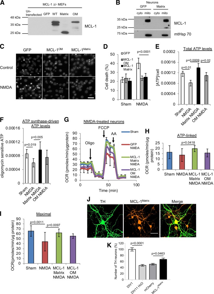
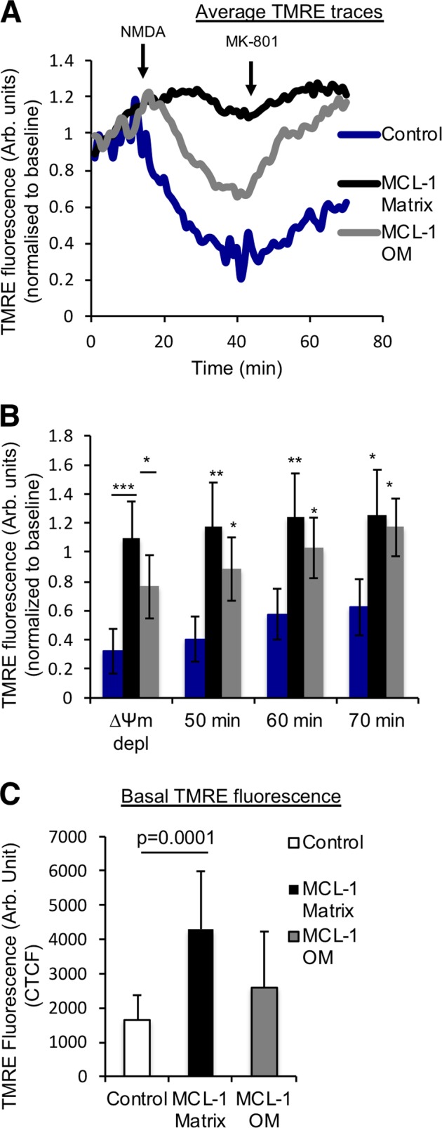
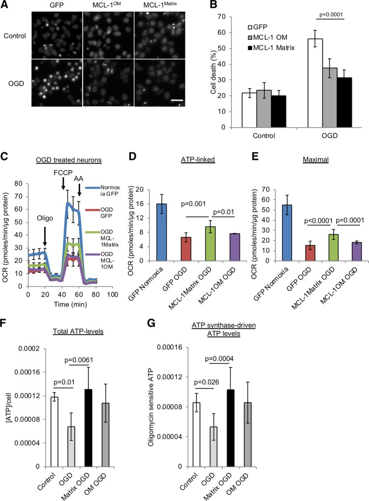
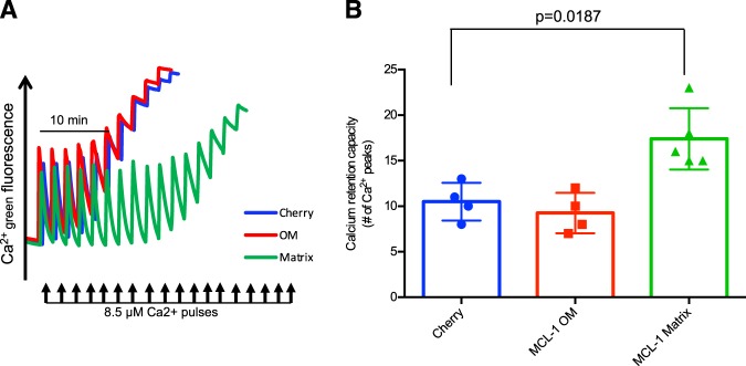
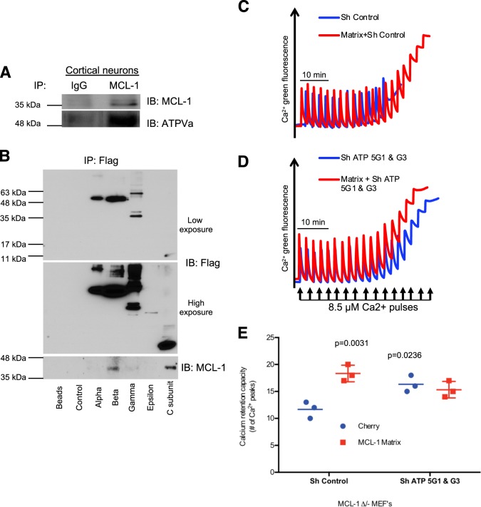
Mitochondria play a crucial role in neuronal survival through efficient energy metabolism. In pathological conditions, mitochondrial stress leads to neuronal death, which is regulated by the anti-apoptotic BCL-2 family of proteins. MCL-1 is an anti-apoptotic BCL-2 protein localized to mitochondria either in the outer membrane (OM) or inner membrane (Matrix), which have distinct roles in inhibiting apoptosis and promoting bioenergetics, respectively. While the anti-apoptotic role for Mcl1 is well characterized, the protective function of MCL-1 Matrix remains poorly understood. Here, we show MCL-1OM and MCL-1Matrix prevent neuronal death through distinct mechanisms. We report that MCL-1Matrix functions to preserve mitochondrial energy transduction and improves respiratory chain capacity by modulating mitochondrial oxygen consumption in response to mitochondrial stress. We show that MCL-1Matrix protects neurons from stress by enhancing respiratory function, and by inhibiting mitochondrial permeability transition pore opening. Taken together, our results provide novel insight into how MCL-1Matrix may confer neuroprotection under stress conditions involving loss of mitochondrial function.


Conflict of interest statement

The authors declare that they have no conflict of interest.

Figures

Fig. 1
Fig. 1. MCL-1 protects neurons against mild mitochondrial stress induced by NMDA excitotoxicity and promotes survival of Parkin KO dopamine neurons.
a Assessment of MCL-1 expression pattern in MCL-1 ∆/- MEFs transduced with lentivirus carrying GFP, MCL-1 WT, MCL-1Matrix, and MCL-1OM. b Immunoblots from cytosolic fraction (cyto) and mitochondrial-enriched fraction (mito) in WT cortical neurons expressing GFP or MCL-1Matrix. Mitochondrial Hsp70 was observed only in mitochondrial fraction. Representative Hoechst images showing healthy (uniformly labeled) and dead (condensed) nuclei in cortical neurons expressing GFP, MCL-1Matrix or MCL-1OM in response to NMDA excitation 24 h post treatment. Scale bar 20 µm. c Cortical neurons were transduced as indicated and cell death was quantified in response to NMDA excitation 24 h post treatment (averages ± SD of nine replicates from three independent experiments). d Total ATP levels in cortical neurons expressing GFP, MCL-1Matrix or MCL-1OM in response NMDA excitation 24 h post treatment. e, f ATP in cells subjected to the same conditions as in (e) and treated with oligomycin (10 µM) for 1 h. f Total ATP levels—oligomycin sensitive ATP levels are shown (averages ± SD of nine replicates from three independent experiments). gi Cortical neurons transduced with GFP, MCL-1Matrix or MCL-1OM was treated with NMDA (100 µM/ 10 µM glycine) for 30 min and OCR was measured 24 h post excitation using a Seahorse XF24 Extracellular Flux Analyzer (g). Quantification of ATP-linked (baseline OCR minus oligomycin-insensitive OCR) (h) and Maximal respiration capacity (FCCP-induced OCR) (i) (averages ± SD of nine replicates from three independent experiments). j Representative images of primary mouse substantia nigra dopamine neurons from Parkin KO mouse showing tyrosine hydroxylase (TH) in green, MCL-1Matrix co-expressing cherry (red) and overlay of both images. k Dopamine neurons were transduced as indicated and survival was quantified at 11 days in vitro (DIV) compared to the number detected at DIV 1 after initial plating to monitor the extent of spontaneous neuronal loss in Parkin KO neurons (averages ± SD from 11–13 coverslips). Scale bar 50 µm. Data information: one-way ANOVA followed by Tukey’s post hoc test.
Fig. 2
Fig. 2. MCL-1Matrix maintains mitochondrial membrane potential in response to acute neuronal stress.
a Average TMRE traces as in neurons transduced with lentivirus carrying Control, MCL-1Matrix and MCL-1OM during NMDA excitation. b Quantification of TMRE traces in (a) at the lowest value (∆Ψm depolarization) following NMDA excitation and at different time points as indicated. c Quantification of basal TMRE fluorescence in neurons expressing Control, MCL-1Matrix, and MCL-1OM (average ± SD of 12–14 neurons from two independent experiments). Data information: one-way ANOVA followed by Tukey’s post hoc test. *P < 0.05, **P < 0.01 and ***P < 0.001.
Fig. 3
Fig. 3. MCL-1Matrix protects neurons and enhances mitochondrial bioenergetics in response to oxygen glucose deprivation.
a, b Representative Hoechst images showing healthy (uniformly labeled) and dead (condensed) nuclei in cortical neurons expressing GFP, MCL-1Matrix or MCL-1OM in response to OGD (a) and quantification of cell death is shown in (b) (averages ± SD of nine replicates from three independent experiments). ce Cortical neurons expressing GFP, MCL-1Matrix, and MCL-1OM was treated with either normoxia or oxygen glucose deprivation (OGD) for 2 h and OCR was measured 24 h post injury (c). Quantification of ATP-linked (baseline OCR minus oligomycin-insensitive OCR) (d) and Maximal respiration capacity (FCCP-induced OCR) (e) (averages ± SD of 12 replicates from three independent experiments). f, g Total ATP levels (f) and ATP synthase-driven (Total ATP levels—oligomycin sensitive) ATP levels are shown in (g) (averages ± SD of nine replicates from three independent experiment Data information: one-way ANOVA followed by Tukey’s post hoc test.
Fig. 4
Fig. 4. MCL-1Matrix regulates mitochondrial calcium retention capacity.
a, b Calcium retention capacity (CRC) in MCL-1 ∆/- MEFs expressing cherry, MCL-1OM or MCL-1Matrix exposed to consecutive pulses of Ca2+ (8.5 µM). Experiments were performed in the presence of Succinate (5 mM), Rotenone (1 µM) and Pi (10 mM) (a). Quantification of number of calcium pulses before pore opening (b) (average ± SD from four to five independent experiments). Data information: one-way ANOVA followed by Tukey’s post hoc test.
Fig. 5
Fig. 5. MCL-1Matrix regulates Mitochondrial permeability transition pore through functional interactions with ATP synthase.
a Endogenous MCL-1 was immunoprecipitated from cortical neurons lysates and the eluted samples were analyzed for MCL-1 and ATP5A expression by western blot. b Cherry (control), Alpha, Beta, Gamma, Epsilon, and C subunit of ATP synthase constructs were transiently transfected into 293 T cells. Fourty-eight hours post-transfection, cells were lysed; immunoprecipitated with anti-flag beads and analyzed by western blot. c–e) Calcium retention capacity (CRC) in MCL-1 ∆/- MEFs expressing Cherry or MCL-1Matrix and co-transduced with either ShControl or ShATP5G1 & G3. e Quantification of CRC shown in (c, d) (average ± SD from three independent experiments). Data information: one-way ANOVA followed by Tukey’s post hoc test.

References

    1. Krieger C, Duchen MR. Mitochondria, Ca2+ and neurodegenerative disease. Eur. J. Pharmacol. 2002;447:177–188. doi: 10.1016/S0014-2999(02)01842-3. - DOI - PubMed
    1. Duchen MR. Mitochondria, calcium-dependent neuronal death and neurodegenerative disease. Pflug. Arch. 2012;464:111–121. doi: 10.1007/s00424-012-1112-0. - DOI - PMC - PubMed
    1. Nicholls DG. Mitochondrial calcium function and dysfunction in the central nervous system. Biochim. Biophys. Acta. 2009;1787:1416–1424. doi: 10.1016/j.bbabio.2009.03.010. - DOI - PMC - PubMed
    1. Bakthavachalam P, Shanmugam PST. Mitochondrial dysfunction - Silent killer in cerebral ischemia. J. Neurol. Sci. 2017;375:417–423. doi: 10.1016/j.jns.2017.02.043. - DOI - PubMed
    1. Manfredi G, Xu Z. Mitochondrial dysfunction and its role in motor neuron degeneration in ALS. Mitochondrion. 2005;5:77–87. doi: 10.1016/j.mito.2005.01.002. - DOI - PubMed

Publication types

MeSH terms

Substances