. 2020 May 5;10(1):7538.
doi: 10.1038/s41598-020-64437-y.

The F220C and F45L rhodopsin mutations identified in retinitis pigmentosa patients do not cause pathology in mice

Affiliations

The F220C and F45L rhodopsin mutations identified in retinitis pigmentosa patients do not cause pathology in mice

Tylor R Lewis et al. Sci Rep. .

Abstract

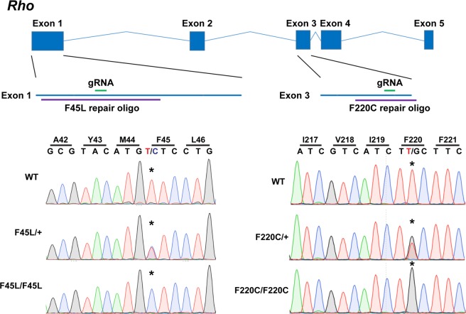
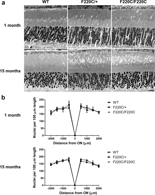
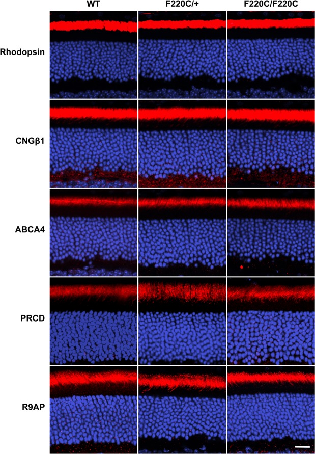
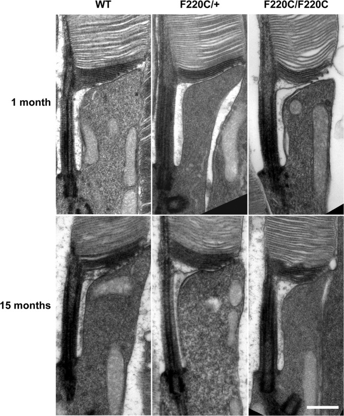
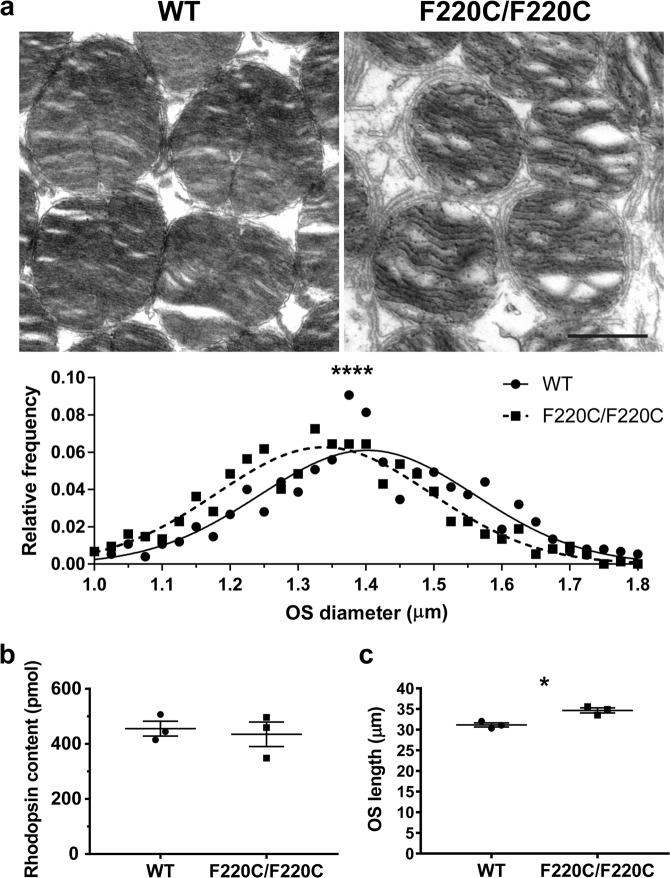
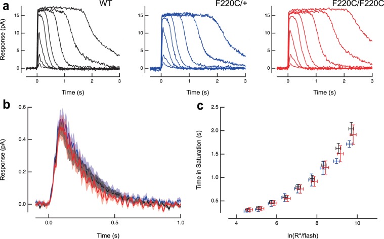
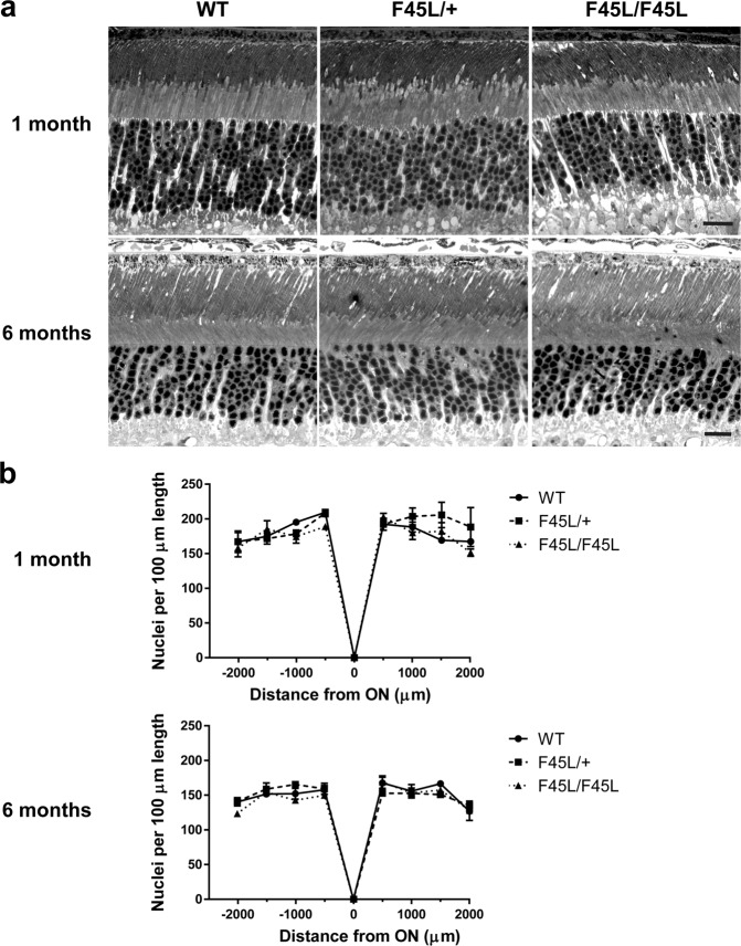
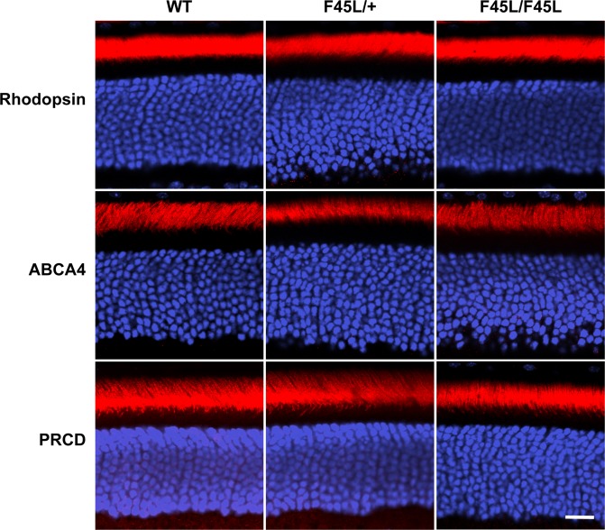
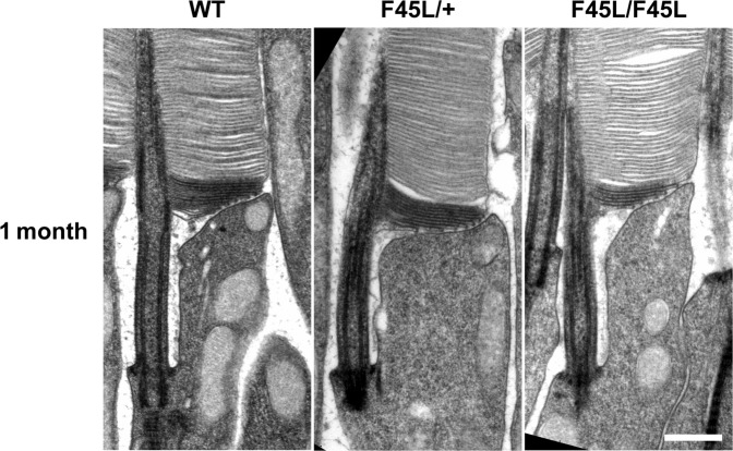
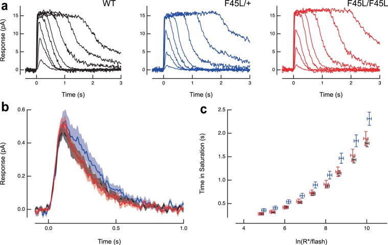
Retinitis pigmentosa is a retinal degenerative disease that leads to blindness through photoreceptor loss. Rhodopsin is the most frequently mutated protein in this disease. While many rhodopsin mutations have well-understood consequences that lead to cell death, the disease association of several rhodopsin mutations identified in retinitis pigmentosa patients, including F220C and F45L, has been disputed. In this study, we generated two knockin mouse lines bearing each of these mutations. We did not observe any photoreceptor degeneration in either heterozygous or homozygous animals of either line. F220C mice exhibited minor disruptions of photoreceptor outer segment dimensions without any mislocalization of outer segment proteins, whereas photoreceptors of F45L mice were normal. Suction electrode recordings from individual photoreceptors of both mutant lines showed normal flash sensitivity and photoresponse kinetics. Taken together, these data suggest that neither the F220C nor F45L mutation has pathological consequences in mice and, therefore, may not be causative of retinitis pigmentosa in humans.


Conflict of interest statement

The authors declare no competing interests.

Figures

Figure 1
Figure 1
Generation of knockin mouse lines bearing the F220C and F45L rhodopsin mutations. The coding sequence of mouse Rho consists of five exons. (Left) The F45L mutation is caused by a c.133 T > C mutation in exon 1. A 200-nt long repair oligonucleotide (oligo) was used (purple) in conjunction with the depicted (in green) single small guide RNA (gRNA) and Cas9 mRNA and protein to generate this allele. (Right) The F220C mutation is caused by a c.659 T > G mutation in exon 3. A 102-nt long repair oligonucleotide (oligo) was used (purple) in conjunction with the depicted (in green) single small guide RNA (gRNA) and Cas9 protein to generate this allele. Sequencing of DNA from WT, heterozygous, and homozygous mice reveals the correct knockin mutation generated for both the F45L and F220C alleles.
Figure 2
Figure 2
F220C mice do not exhibit photoreceptor degeneration. (a) Light microscopy images of 0.5 μm thin retinal plastic sections stained with methylene blue. Depicted are WT, heterozygous (F220C/+), and homozygous (F220C/F220C) retinas at 1 month and 15 months of age. Scale bar is 10 μm. (b) The number of photoreceptor nuclei are quantified over a 100 μm length of retina at 500 μm intervals away from the optic nerve (ON) at 1 month and 15 months of age (n = 3 for each genotype at each age). There are no statistically significant differences among genotypes with two-way ANOVA at either age.
Figure 3
Figure 3
F220C mice do not exhibit mislocalization of photoreceptor outer segment proteins. Immunofluorescent staining of photoreceptor outer segment-specific proteins in WT, heterozygous (F220C/+), and homozygous (F220C/F220C) retinas at 1 month of age. Both disc-specific (rhodopsin, ABCA4, PRCD, R9AP) and outer segment plasma membrane-specific (CNGβ1) proteins are analyzed (red). Nuclei are stained with Hoechst (blue). Scale bar is 10 μm.
Figure 4
Figure 4
Rod outer segments of F220C mice do not exhibit any gross ultrastructural defects. TEM images of the base of photoreceptor outer segments from WT, heterozygous (F220C/+), and homozygous (F220C/F220C) mice are shown at 1 month and 15 months of age. Sections are stained with tannic acid to intensely label nascent, open discs at the base of the outer segment. Scale bar is 0.5 μm.
Figure 5
Figure 5
Rod outer segments of F220C mice are slightly thinner and longer. (a) TEM of a tangential section through the photoreceptor outer segment layer of WT and homozygous F220C mice. Scale bar is 1 μm. Diameters of 150 outer segments were measured in 5 different mice for a total of 750 outer segments per genotype. Diameters were grouped within 0.02 μm bins to plot the relative frequency of outer segment diameters ranging from 1.0 μm to 1.8 μm. The relative frequency plot is overlaid with Gaussian distribution curves for WT (solid line) and F220C/F220C (dashed line) datasets. Unpaired t-test reveals a statistically significant difference (p < 0.0001) between the outer segment diameters of WT (1.40 ± 0.01 µm) and F220C/F220C (1.34 ± 0.01 µm) mice. (b) Total rhodopsin content of dissected eyecups was determined by difference spectroscopy. Unpaired t-test depicts no statistically significant difference (p = 0.7109) in rhodopsin content between WT and homozygous F220C mice at 10 months of age (n = 3 for each genotype). (c) Photoreceptor outer segment lengths were measured from five different regions of each of three different mice per genotype at 1 month of age. Unpaired t-test reveals a statistically significant difference (p = 0.011) between the outer segment lengths of WT (34.7 ± 0.6 µm) and F220C/F220C (31.1 ± 0.5 µm) mice.
Figure 6
Figure 6
F220C rhodopsin mutant photoreceptors do not exhibit any electrophysiological defects. (a) Suction electrode recordings of families of responses to flashes that ranged from 13–51,000 photons/μm2 by factors of 2–4. Flashes were delivered at t = 0 s. (b) Population average single photon responses calculated from WT (n = 22), F220C/+ (n = 14) and F220C/F220C (n = 11) rods. Light shading represents SEM. (c) Relationship between the time that a bright flash response remained in saturation and the natural log of the number of photoexcited rhodopsins (R*) produced by the flash. The initial slope reflects the dominant time constant of recovery for bright flashes, which was the same for mutant and WT rods. Error bars represent SEM.
Figure 7
Figure 7
F45L mice do not exhibit photoreceptor degeneration. (a) Light microscopy images of 0.5 μm thin retinal plastic sections stained with methylene blue. Depicted are WT, heterozygous (F45L/+), and homozygous (F45L/F45L) retinas at 1 month and 6 months of age. Scale bars are 10 μm. (b) The number of photoreceptor nuclei are quantified over a 100 μm length of retina at 500 μm intervals away from the optic nerve (ON) at 1 month and 6 months of age (n = 3 for each genotype at each age). There are no statistically significant differences among genotypes with two-way ANOVA at either age.
Figure 8
Figure 8
F45L mice do not exhibit mislocalization of photoreceptor outer segment proteins. Immunofluorescent staining of photoreceptor outer segment disc-specific proteins (red) in WT, heterozygous (F45L/+), and homozygous (F45L/F45L) retinas at 1 month of age. Nuclei are stained with Hoechst (blue). Scale bar is 10 μm.
Figure 9
Figure 9
Rod outer segments of F45L mice do not exhibit any gross ultrastructural defects. TEM images of the base of photoreceptor outer segments from WT, heterozygous (F45L/+), and homozygous (F45L/F45L) mice are shown at 1 month of age. Sections are stained with tannic acid to intensely label nascent, open discs at the base of the outer segment. Scale bar is 0.5 μm.
Figure 10
Figure 10
F45L rhodopsin mutant photoreceptors do not exhibit any electrophysiological defects. (a) Suction electrode recordings of families of responses to flashes that ranged from 6–50,000 photons/μm2 by factors of 2–4. Flashes were delivered at t = 0 s. (b) Population average single photon responses calculated from WT (n = 22), F45L/+ (n = 14) and F45L/F45L (n = 19) rods. Light shading represents SEM. (c) Relationship between the time that a bright flash response remained in saturation and the natural log of the number of photoexcited rhodopsins (R*) produced by the flash. The initial slope reflects the dominant time constant of recovery for bright flashes, which was the same for mutant and WT rods. Error bars represent SEM.

Similar articles

Cited by

References

    1. Farrar GJ, Kenna PF, Humphries P. On the genetics of retinitis pigmentosa and on mutation-independent approaches to therapeutic intervention. Embo j. 2002;21:857–864. doi: 10.1093/emboj/21.5.857. - DOI - PMC - PubMed
    1. Hartong DT, Berson EL, Dryja TP. Retinitis pigmentosa. Lancet. 2006;368:1795–1809. doi: 10.1016/s0140-6736(06)69740-7. - DOI - PubMed
    1. Stenson PD, et al. The Human Gene Mutation Database: building a comprehensive mutation repository for clinical and molecular genetics, diagnostic testing and personalized genomic medicine. Hum Genet. 2014;133:1–9. doi: 10.1007/s00439-013-1358-4. - DOI - PMC - PubMed
    1. Mendes HF, van der Spuy J, Chapple JP, Cheetham ME. Mechanisms of cell death in rhodopsin retinitis pigmentosa: implications for therapy. Trends Mol Med. 2005;11:177–185. doi: 10.1016/j.molmed.2005.02.007. - DOI - PubMed
    1. Bunge S, et al. Molecular analysis and genetic mapping of the rhodopsin gene in families with autosomal dominant retinitis pigmentosa. Genomics. 1993;17:230–233. doi: 10.1006/geno.1993.1309. - DOI - PubMed

Publication types