. 2020 May 5;3(1):209.
doi: 10.1038/s42003-020-0927-z.

The coenzyme thiamine diphosphate displays a daily rhythm in the Arabidopsis nucleus

Affiliations

The coenzyme thiamine diphosphate displays a daily rhythm in the Arabidopsis nucleus

Zeenat B Noordally et al. Commun Biol. .

Abstract

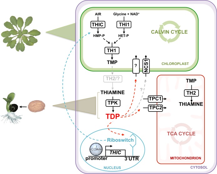
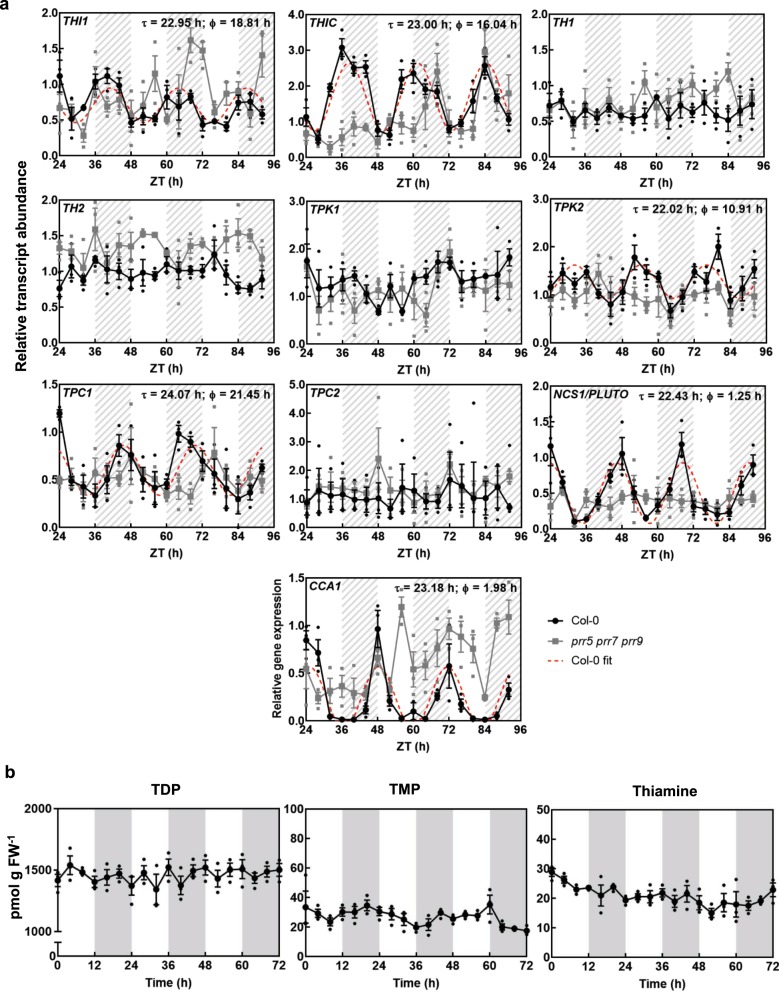
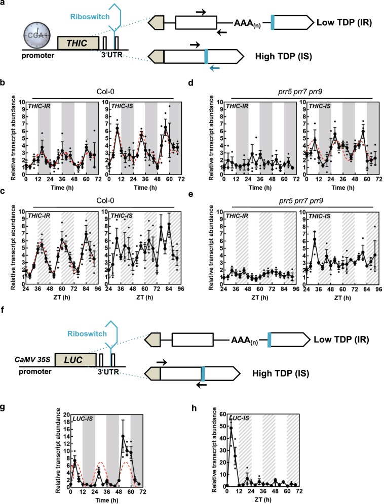
In plants, metabolic homeostasis-the driving force of growth and development-is achieved through the dynamic behavior of a network of enzymes, many of which depend on coenzymes for activity. The circadian clock is established to influence coordination of supply and demand of metabolites. Metabolic oscillations independent of the circadian clock, particularly at the subcellular level is unexplored. Here, we reveal a metabolic rhythm of the essential coenzyme thiamine diphosphate (TDP) in the Arabidopsis nucleus. We show there is temporal separation of the clock control of cellular biosynthesis and transport of TDP at the transcriptional level. Taking advantage of the sole reported riboswitch metabolite sensor in plants, we show that TDP oscillates in the nucleus. This oscillation is a function of a light-dark cycle and is independent of circadian clock control. The findings are important to understand plant fitness in terms of metabolite rhythms.


Conflict of interest statement

The authors declare no competing interests.

Figures

Fig. 1
Fig. 1. Overview of thiamine metabolism in plants.
Thiamine monophosphate (TMP) is biosynthesized de novo in the chloroplast by condensation of hydroxymethylpyrimidine-phosphate (HMP-P) and hydroxyethylthiazole-phosphate (HET-P), which are derived from 5-aminoimidazole ribotide (AIR), nicotinamide adenine dinucleotide (NAD+) and glycine as precursors, followed by dephosphorylation and pyrophosphorylation to thiamine diphosphate (TDP) in the cytosol. The pathway involves the enzyme action of THIC, THI1, TH1, TPK and TH2, although the latter enzyme appears to be predominantly in the mitochondrion. TDP is needed for central metabolic pathways in organelles: the TCA cycle and the Calvin cycle, as well as glycolysis in the cytoplasm (not shown). While TPCs (TPC1 and TPC2) import TDP at the mitochondrial membrane, it is not known how TDP is transported into the chloroplast or nonphotosynthetic plastids, although the promiscuous transporter NCS1 is thought to import HMP or TDP (represented by the gray arrow and by “?”). In the nucleus, expression of the THIC gene is regulated by the circadian clock through a CCA1 binding site in its promoter and by the level of free TDP through a riboswitch present in the 3-UTR. The biosynthesis de novo pathway of TDP is active in green tissue about 5 days after germination (indicated by a green triangle), whereas TDP is supplied through the action of TPK on thiamine in germinating seeds (indicated by a brown triangle). The blue arrow represents negative feedback regulation of HMP-P production caused by the binding of TDP to the THIC riboswitch in the nucleus. The red arrows represent the necessity to transport TDP into the organelles.
Fig. 2
Fig. 2. Central metabolism depends on thiamine supply.
a Photographs of 11-day-old wild type and thiC Arabidopsis seedlings supplemented with thiamine levels as indicated. Seedlings were grown on 1/2 MS agar plates under a 16-h photoperiod (120 μmol photons m−2 s−1) at 22 °C and 8 h of darkness at 18 °C. b Levels of thiamine diphosphate (TDP) measured in seedlings of wild type and the thiC mutant as shown in (a) as a function of the thiamine supplementation level as indicated. Data of three individual biological replicates of pooled material (n = 15) is shown with error bars representing SE. c Significant metabolite content changes as a function of thiamine supplementation (as in (a)) using nontargeted metabolomics. Sphere size and color shade correlate with the extent of change of the particular metabolite as indicated. The plot reports all metabolites out of 372 detected that were found to pass significance criteria (|log2(fold-change)| > 0.5 and adj. p value < 0.01) in at least one condition. The data shown are relative to the control with no thiamine supplementation. Significant points are encircled in gray.
Fig. 3
Fig. 3. The plastid localized NCS1/PLUTO is predominant in root tissue.
a Representative pictures of Arabidopsis mesophyll protoplasts expressing the NCS1/PLUTO-YFP fusion protein compared to YFP alone. The scale bar represents 10 μm. b Cartoon of the predicted topology of NCS1/PLUTO as determined by TMpred (http://www.ch.embnet.org/software/TMPRED_form.html). The numbers refer to the corresponding amino acid residues. c Representative images of histochemical analysis of Arabidopsis expressing GUS under the control of the promoter of NCS1/PLUTO: 7-d-old seedling, differentiation zone of a mature root and a developing silique, 7-d-old seedling root tip, mature rosette and a flower. The scale bars represent 0.1 cm. d Representative pictures of tissue expression of NCS1/PLUTO-YFP compared to YFP alone in stable transformants of Arabidopsis either by confocal microscopy of roots (columns 1 and 2, the scale bar represents 100 μm) or epifluorescence of young seedlings and roots (column 3, the scale bar represents 0.2 cm). e Tissue expression analysis of NCS1/PLUTO transcript levels by qPCR. Data of three individual biological replicates is shown with error bars representing SE. Transcript levels are relative to PDF2 (At1g13320) and each sample was referenced to siliques (set to 1). Plants were either grown on 1/2 MS agar plates or on soil under a 16-h photoperiod (120 μmol photons m−2 s−1) at 22 °C and 8 h of darkness at 18 °C.
Fig. 4
Fig. 4. NCS1/PLUTO is required for plant development during compromised TDP biosynthesis de novo.
a Gene model of NCS1/PLUTO with the single exon depicted as a black bar. The location of the T-DNA insertions in WISCDSLOX419C03 (ncs1-1) as depicted were confirmed by genotyping and sequencing. b Quantitative analysis of NCS1/PLUTO expression in ncs1-1 relative to wild type (Col-0) from 11-day-old seedlings. Data of three individual biological replicates is shown with error bars representing SE. The asterisk indicates a significant difference as a result of a t test (p ≤ 0.01). Transcript levels are relative to PDF2 (At1g13320). c Representative photographs of 13-day-old ncs1-1, thiC and ncs1-1 thiC (the numbers refer to three independent crosses) mutant lines in the absence (0) or presence of thiamine supplementation levels as indicated compared to wild type (Col-0). Seedlings were grown on 1/2 MS agar plates under a 16-h photoperiod (120 μmol photons m−2 s−1) at 22 °C and 8 h of darkness at 18 °C.
Fig. 5
Fig. 5. Temporal separation of TDP biosynthesis and transport by the circadian clock.
a Transcript abundance of TDP biosynthesis and transport genes by qPCR in continuous light (free-running conditions) in wild type (Col-0, black) compared to the arrhythmic triple-mutant prr5 prr7 prr9 (gray). The dashed lines correspond to cases where cosine waves could be fitted to the data using the FFT-NLLS method from BioDare2 with the estimated period (Φ) and phase (τ) as indicated. Transcript levels are relative to UBC21 (At5g25760) and each sample was referenced to wild-type ZT 24. The transcript profile of CCA1 under the same conditions is shown as a control. Plants were grown in culture on 1/2 MS agar plates and entrained for 13 days in equinoctial conditions (12-h photoperiod with 120 μmol photons m−2 s−1 and 12 h of darkness at 20 °C) before transfer to constant light after which shoot material was harvested from seedlings (n = 10) every 4 h at the ZT times indicated. White and hatched gray areas represent subjective day and night, respectively. Data of three individual biological replicates are shown with error bars representing SE. b Thiamine, thiamine monophosphate (TMP) or thiamine diphosphate (TDP) content of wild type (Col-0), as determined by HPLC from shoot material of 14-day-old seedlings grown in culture on 1/2 MS agar plates under equinoctial conditions (12-h photoperiod with 120 μmol photons m−2 s−1 and 12 h of darkness at 20 °C). Data of three individual biological replicates of pooled material (n = 20−25) is shown with error bars representing SE.
Fig. 6
Fig. 6. Nuclear TDP levels oscillate in a circadian clock independent manner.
a Gene model of THIC emphasizing splice variants of the 3-UTR as a function of TDP levels. Exons are depicted as white/beige boxes, introns as lines and the polyadenylation (AAA(n)) tail is as indicated. CCA1 binds in the promoter region of THIC. The TDP riboswitch (blue) is at the end of the second intron in the 3-UTR. When TDP levels are low the intron is retained (IR), whereas the intron is spliced (IS) when the TDP level is high. The arrows indicate primer-binding sites for monitoring IS and IR levels. b, c Transcript abundance of IR or IS variants of THIC (THIC-IR or THIC-IS, respectively) by qPCR in equinoctial light/dark cycles (b) or continuous light (c) in wild type (Col-0). White and dark gray areas represent light and dark in equinoctial conditions, whereas white and hatched gray areas represent subjective day and night in continuous light conditions. Data of three individual biological replicates are shown with error bars representing SE. The red dashed lines correspond to cases where cosine waves could be fitted to the data using the FFT-NLLS method from BioDare2. d, e As for c, d but in the arrhythmic triple-mutant prr5 prr7 prr9. f Gene model of the LUCIFERASE (LUC) construct expressed under the control of the CaMV 35S promoter and with the THIC 3-UTR. Alternative splicing is as for a and is dependent on TDP levels in the nucleus. Arrows indicate primer-binding sites in the 3-UTR for monitoring IS levels (LUC-IS). g, h Transcript abundance of IS variants of LUC (LUC-IS) by qPCR in either equinoctial light/dark cycles (g) or continuous light (h) in wild type. Data of three individual biological replicates are shown with error bars representing SE. The red dashed line corresponds to cosine wave fitting of the data using the FFT-NLLS method from BioDare2. In all cases, plants were grown in culture on 1/2 MS agar plates and entrained for 13 days in equinoctial conditions (12-h photoperiod with 120 μmol photons m−2 s−1 and 12 h of darkness at 20 °C) and either transferred to constant light or retained in equinoctial conditions. Shoot material was harvested over 3 days at 4-h intervals from seedlings (n = 10) at the times indicated. Transcript levels are relative to UBC21 (At5g25760).

References

    1. Bunik VI, Tylicki A, Lukashev NV. Thiamin diphosphate-dependent enzymes: from enzymology to metabolic regulation, drug design and disease models. FEBS J. 2013;280:6412–6442. doi: 10.1111/febs.12512. - DOI - PubMed
    1. Goyer A. Thiamine in plants: aspects of its metabolism and functions. Phytochemistry. 2010;71:1615–1624. doi: 10.1016/j.phytochem.2010.06.022. - DOI - PubMed
    1. Lichtenthaler HK. Biosynthesis, accumulation and emission of carotenoids, alpha-tocopherol, plastoquinone, and isoprene in leaves under high photosynthetic irradiance. Photosynth. Res. 2007;92:163–179. doi: 10.1007/s11120-007-9204-y. - DOI - PubMed
    1. Chatterjee A, et al. Saccharomyces cerevisiae THI4p is a suicide thiamine thiazole synthase. Nature. 2011;478:542–546. doi: 10.1038/nature10503. - DOI - PMC - PubMed
    1. Garcia AF, et al. THI1, a protein involved in the biosynthesis of thiamin in Arabidopsis thaliana: structural analysis of THI1(A140V) mutant. Biochim. Biophys. Acta. 2014;1844:1094–1103. doi: 10.1016/j.bbapap.2014.03.005. - DOI - PubMed

Publication types

Substances