Nonlinear optical microscopy is a novel tool for the analysis of cutaneous alterations in pseudoxanthoma elasticum
- PMID: 32372237
- PMCID: PMC7505829
- DOI: 10.1007/s10103-020-03027-w
Nonlinear optical microscopy is a novel tool for the analysis of cutaneous alterations in pseudoxanthoma elasticum
Abstract
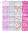
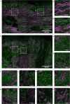
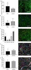
Pseudoxanthoma elasticum (PXE, OMIM 264800) is a rare autosomal recessive disorder with ectopic mineralization and fragmentation of elastin fibers. It is caused by mutations of the ABCC6 gene that leads to decreased serum levels of inorganic pyrophosphate (PPi) anti-mineralization factor. The occurrence of severe complications among PXE patients highlights the importance of early diagnosis so that prompt multidisciplinary care can be provided to patients. We aimed to examine dermal connective tissue with nonlinear optical (NLO) techniques, as collagen emits second-harmonic generation (SHG) signal, while elastin can be excited by two-photon excitation fluorescence (TPF). We performed molecular genetic analysis, ophthalmological and cardiovascular assessment, plasma PPi measurement, conventional histopathological examination, and ex vivo SHG and TPF imaging in five patients with PXE and five age- and gender-matched healthy controls. Pathological mutations including one new variant were found in the ABCC6 gene in all PXE patients and their plasma PPi level was significantly lower compared with controls. Degradation and mineralization of elastin fibers and extensive calcium deposition in the mid-dermis was visualized and quantified together with the alterations of the collagen structure in PXE. Our data suggests that NLO provides high-resolution imaging of the specific histopathological features of PXE-affected skin. In vivo NLO may be a promising tool in the assessment of PXE, promoting early diagnosis and follow-up.
Keywords: Calcification; Elastin; Multiphoton microscopy; Nonlinear optical microscopy; Pseudoxanthoma elasticum.
Conflict of interest statement
The authors declare that they have no conflict of interest.
Figures


References
-
- Jansen RS, Duijst S, Mahakena S, Sommer D, Szeri F, Váradi A, Plomp A, Bergen AA, Oude Elferink RP, Borst P. ABCC6–mediated ATP secretion by the liver is the main source of the mineralization inhibitor inorganic pyrophosphate in the systemic circulation—brief report. Arterioscler Thromb Vasc Biol. 2014;34:1985–1989. doi: 10.1161/ATVBAHA.114.304017. - DOI - PMC - PubMed
-
- Bäck M, Aranyi T, Cancela ML, Carracedo M, Conceição N, Leftheriotis G, Macrae V, Martin L, Nitschke Y, Pasch A. Endogenous calcification inhibitors in the prevention of vascular calcification: a consensus statement from the COST action EuroSoftCalcNet. Front Cardiovasc Med. 2019;5:196. doi: 10.3389/fcvm.2018.00196. - DOI - PMC - PubMed
MeSH terms
Substances
Grants and funding
- ÚNKP-19-3-II-SE-15/New National Excellence Program of the Ministry for Innovation and Technology
- ÚNKP-19-3-I-SE-78/New National Excellence Program of the Ministry for Innovation and Technology
- ÚNKP-18-3-I-SE-65/New National Excellence Program of the Ministry for Innovation and Technology
- K-129047, 2018/National Research, Development and Innovation Fund of Hungary
- NKM-93/2018/Hungarian Academy of Sciences
- János Bolyai Scholarship/Hungarian Academy of Sciences
- EFOP-3.6.3-VEKOP-16-2017-00009/Kiegészítő Kutatási Kiválósági Ösztöndíj
- FK_131916, 2019/National Research, Development and Innovation Fund of Hungary
- COST action CA16115/EuroSoftCalcNet
- Mobility grant from the Hungarian Academy of Sciences/Mobility grant from the Hungarian Academy of Sciences
LinkOut - more resources
Full Text Sources

