. 2020 Oct;17(4):2041-2053.
doi: 10.1007/s13311-020-00865-y.

Cerebellar Astrocyte Transduction as Gene Therapy for Megalencephalic Leukoencephalopathy

Affiliations

Cerebellar Astrocyte Transduction as Gene Therapy for Megalencephalic Leukoencephalopathy

Angela Sánchez et al. Neurotherapeutics. 2020 Oct.

Abstract

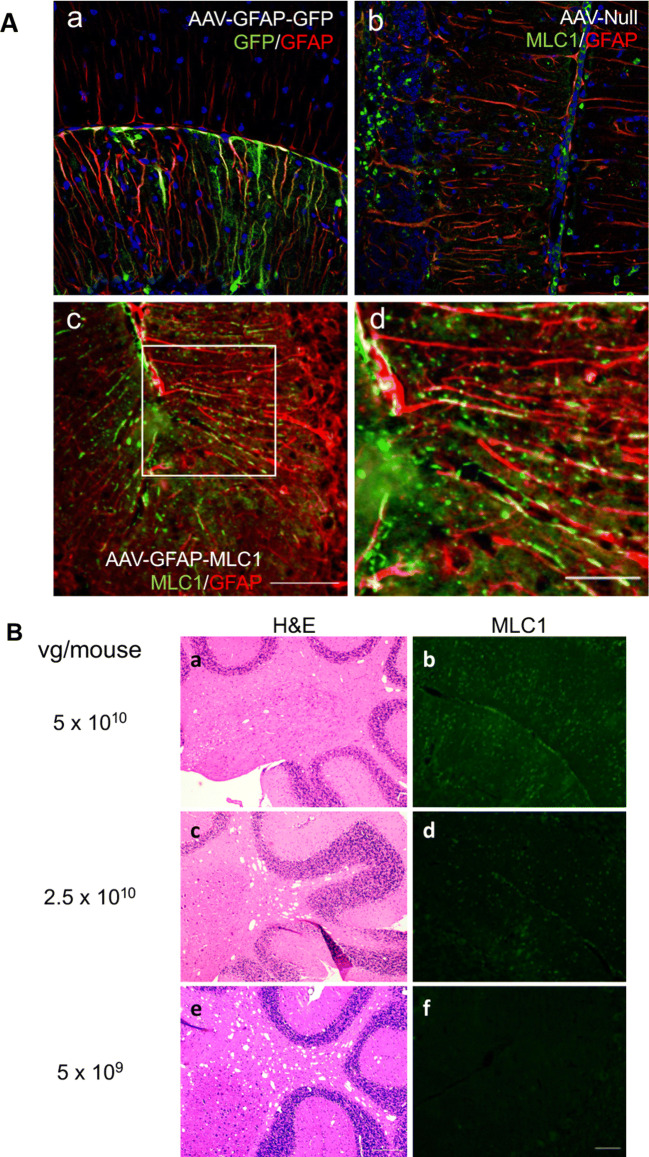
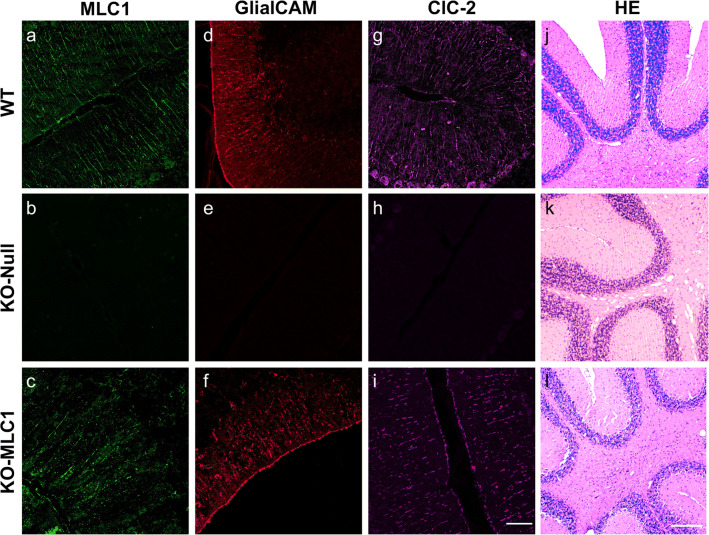
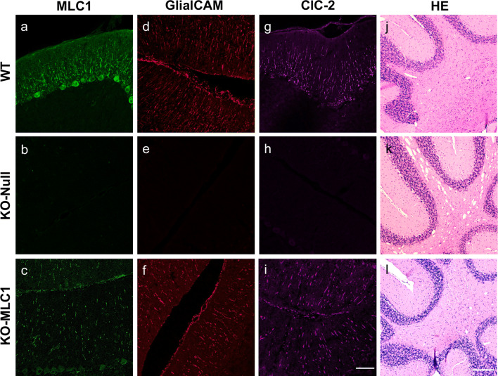
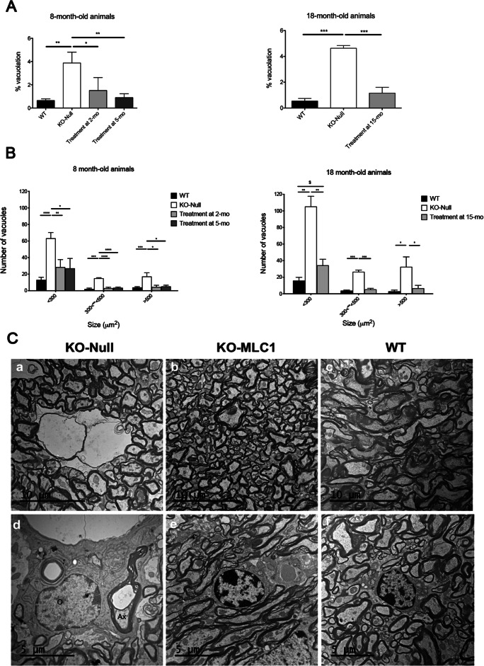
Megalencephalic leukoencephalopathy with subcortical cysts (MLC) is a rare genetic disorder belonging to the group of vacuolating leukodystrophies. It is characterized by megalencephaly, loss of motor functions, epilepsy, and mild mental decline. In brain biopsies of MLC patients, vacuoles were observed in myelin and in astrocytes surrounding blood vessels. There is no therapy for MLC patients, only supportive treatment. We show here a preclinical gene therapy approach for MLC using the Mlc1 knock-out mouse. An adeno-associated virus coding for human MLC1 under the control of the glial fibrillary acidic protein promoter was injected in the cerebellar subarachnoid space of Mlc1 knock-out and wild-type animals at 2 months of age, before the onset of the disease, as a preventive approach. We also tested a therapeutic strategy by injecting the animals at 5 months, once the histopathological abnormalities are starting, or at 15 months, when they have progressed to a more severe pathology. MLC1 expression in the cerebellum restored the adhesion molecule GlialCAM and the chloride channel ClC-2 localization in Bergmann glia, which both are mislocalized in Mlc1 knock-out model. More importantly, myelin vacuolation was extremely reduced in treated mice at all ages and correlated with the amount of expressed MLC1 in Bergmann glia, indicating not only the preventive potential of this strategy but also its therapeutic capacity. In summary, here we provide the first therapeutic approach for patients affected with MLC. This work may have also implications to treat other diseases affecting motor function such as ataxias.

Keywords: AAVrh10; Gene therapy; GlialCAM; MLC; cerebellum; myelin.


Figures

Fig. 1
Fig. 1
Intracerebellar administration in the subarachnoid space is the most efficient route to transduce Bergmann glia. (A) Scheme of the viral construct containing GFP as reporter gene under the regulation of the GFAP promoter. (B) Representation of the five administration routes tested in this study. (C) Images were taken using a fluorescence microscope and showed direct GFP fluorescence in the cerebellum of WT animals injected with the AAV-GFAP-GFP by lumbar intrathecal (IT) administration, by intracerebroventricular (ICV) injection, and by intra cerebellar delivery in the white matter (ICB WM), in the molecular layer (ICB ML), or in the subarachnoid space (ICB SB). Scale bar: 100 μm
Fig. 2
Fig. 2
(A) Subarachnoid administration in the cerebellum of the AAV-GFAP-MLC1 vector in Mlc1 KO animals recovers the expression of MLC1 in Bergmann glia. (a) GFP expression in Bergmann glia in the cerebellum of Mlc1 KO animals injected with AAV-GFAP-GFP vector, in green, colocalizes with GFAP astrocytic marker (in red). (b) Undetectable MLC1 expression (in green) in Mlc1 KO animals injected with the AAV-null vector. GFAP is shown in red. (c) Human MLC1 signal, in green, is detected in Bergmann glia stained in red by GFAP marker after AAV-GFAP-MLC1 administration (scale bar, 50 μm). (d) Inset magnification from c, showing colocalization between MLC1 and GFAP immunoreactivity (scale bar 25 μm). Hoescht co-staining for nuclei is in blue. (B) Correlation between AAV-mediated MLC1 levels and white matter vacuolation. Hematoxylin and eosin staining (left) and consecutive slices with MLC1 immunohistochemistry (in green, right) of the cerebellum from animals injected with different doses of vector. There is a direct correlation between the amount of virus, the immunoreactivity of MLC1 in Bergmann glia, and the number of vacuoles in the white matter of the cerebella. Scale bar: 50 μm in dark-field and 200 μm in bright-field images, respectively
Fig. 3
Fig. 3
Experimental design for the gene therapy MLC approach. (A) Scheme of the viral construct containing human MLC1 as therapeutic gene, under the regulation of the GFAP promoter. (B) Three preclinical studies were designed, the first as a preventive strategy where young non-affected Mlc1 KO were treated at 2 months and euthanized 6 months later; and two therapeutic studies with animals treated at 5 (early) and 15 months (late) and analyzed 3 months later
Fig. 4
Fig. 4
Human MLC1 transgene expression before disease onset prevents the development of cerebellum vacuolation in Mlc1 KO animals. (a, d, g) Confocal microscope representative images from WT animals (injected with an AAV-null; n = 4) showing MLC1 (a, green), GlialCAM (d, red), or ClC-2 (g, magenta) immunoreactivity. (b, e, h) Confocal microscope representative images from untreated Mlc1 KO animals (injected with an AAV-Null; n = 3) labeled for MLC1 (b, green), GlialCAM (e, red), or ClC-2 (h, magenta). (c, f, i) Immunofluorescence representative images from AAV-GFAP-MLC1-injected KO animals (n = 4) labeled for MLC1 (c, green), GlialCAM (f, red), or Clc-2 (i, magenta). Absence of MLC1 and mislocalization of GlialCAM and ClC-2 proteins in Mlc1 KO animals is evident, and localization of both proteins is restored by the administration of the AAV-GFAP-MLC1 vector. (j, k, l) Representative bright-field microscope images from hematoxylin-eosin staining of untreated WT (j), untreated Mlc1 KO (k), or treated Mlc1 KO (l) animals showing a significant reduction of the myelin vacuolation in treated animals, similar to WT. Scale bar: 50 μm in confocal microscopy images and 200 μm in bright-field images
Fig. 5
Fig. 5
Human MLC1 administration in disease-affected Mlc1 KO animals at 5 months of age reduces the vacuolation in cerebellar white matter. (a, d, g) Representative images from WT animals (injected with an AAV-Null; n = 4) showing MLC1 (a, green), GlialCAM (d, red), or ClC-2 (g, magenta) immunofluorescence. (b, e, h) Representative fluorescence images from untreated Mlc1 KO animals (injected with an AAV-null; n = 3) showing no immunoreactivity to MLC1 (b, green), GlialCAM (e, red), or ClC-2 (h, magenta). (c, f, i) Representative images from AAV-GFAP-MLC1-injected KO animals (n = 3) showing MLC1 (c, green), GlialCAM (f, red), or ClC-2 immunoreactivity (i, magenta). Results indicated a restoration of MLC1, GlialCAM, and ClC-2 localization by the administration of the AAV-GFAP-MLC1 vector in 5-month-old animals similarly to what was observed when animals were treated at 2 months of age. (j, k, l) Representative images from hematoxylin-eosin staining in paraffin sections of WT (j), untreated Mlc1 KO (k), or treated Mlc1 KO (l) animals show a significant reduction of the myelin vacuolation in treated animals, as occurred with 2-month-old animals. Scale bar: 50 μm in confocal microscopy images and 200 μm in bright-field images
Fig. 6
Fig. 6
MLC1 administration in long-term affected Mlc1 KO mice also reverts the vacuolization phenotype. (a, d, g) Representative images from WT animals (injected with AAV-Null; n = 3) showing MLC1 (a, green), GlialCAM (d, red), or ClC-2 (g, magenta). (b, e, h) Representative immunohistochemistry of samples from untreated Mlc1 KO animals (injected with AAV-null; n = 4) showing MLC1 (b, green), GlialCAM (e, red), or ClC-2 (h, magenta) immunoreactivity. (c, f, i) Representative images from AAV-GFAP-MLC1-injected Mlc1 KO animals (n = 5) showing MLC1 (c, green), GlialCAM (f, red), or ClC-2 (i, magenta). (j, k, l) Hematoxylin-eosin staining in paraffin sections of untreated WT (j), Mlc1 KO (k), or treated Mlc1 KO (l) animals. Scale bar: 50 μm in confocal microscopy images and 200 μm in bright-field images
Fig. 7
Fig. 7
Quantificative and qualitative study of vacuoles in the white matter of the cerebellum. (A) Percentage of vacuolation was quantified in cerebellar sections separated by 40 μm. Values are expressed as percentage of vacuolated area compared to the total area of the white matter in each section in 8- and 18-month-old animals, WT, treated, or untreated Mlc1 KO mice (n = 3 per age and treatment). One-way ANOVA and Tukey post hoc analysis for each time point are depicted on the graph (*p < 0.05; **p < 0.01). (B) Histogram distribution of vacuoles size (in μm2) in WT, treated, or untreated Mlc1 KO mice at 8 and 18 months of age (n = 3 per age and treatment). Statistics: omnibus analysis by one-way ANOVA with Tukey post hoc tests showed statistical significant differences between WT and KO-null and between treated and untreated KO groups but not between WT and treated cohorts. Multiple t tests for the different size range vacuoles show statistically significant differences for all groups and sizes compared with untreated KO (*p < 0.05; **p < 0.01; ***p < 0.001) or between small vacuoles from 18-month-old treated vs WT mice ($p < 0.05). (C) Ultrastructural analyses of myelin vacuolization by transmission electronic microscopy. Myelin vacuolization at two different magnifications showing large vacuoles of aberrant myelin in Mlc1 KO mice (a, d), and normal ultrastructure in Mlc1 KO mice treated with AAV-GFAP-MLC1 (b, e) and WT animals (c, f). ax, axon; o, oligodendrocyte

Similar articles

Cited by

References

    1. van der Knaap MS, Barth PG, Stroink H, et al. Leukoencephalopathy with swelling and a discrepantly mild clinical course in eight children. Ann Neurol. 1995;37:324–334. - PubMed
    1. Singhal BS, Gursahani RD, Udani VP, et al. Megalencephalic leukodystrophy in an Asian Indian ethnic group. Pediatr Neurol. 1996;14:291–296. - PubMed
    1. Ben-Zeev B, Gross V, Kushnir T, et al. Vacuolating megalencephalic leukoencephalopathy in 12 Israeli patients. J Child Neurol. 2001;16:93–99. - PubMed
    1. Harbord MG, Harden A, Harding B, et al. Megalencephaly with dysmyelination, spasticity, ataxia, seizures and distinctive neurophysiological findings in two siblings. Neuropediatrics. 1990;21:164–168. - PubMed
    1. Leegwater PAJ, Yuan BQ, van der Steen J, et al. Mutations of MLC1 (KIAA0027), encoding a putative membrane protein, cause megalencephalic leukoencephalopathy with subcortical cysts. Am J Hum Genet. 2001;68:831–838. - PMC - PubMed

Publication types

Supplementary concepts