. 2020 Jul;111(7):2310-2324.
doi: 10.1111/cas.14444. Epub 2020 Jun 10.

EHF promotes colorectal carcinoma progression by activating TGF-β1 transcription and canonical TGF-β signaling

Affiliations

EHF promotes colorectal carcinoma progression by activating TGF-β1 transcription and canonical TGF-β signaling

Lan Wang et al. Cancer Sci. 2020 Jul.

Abstract

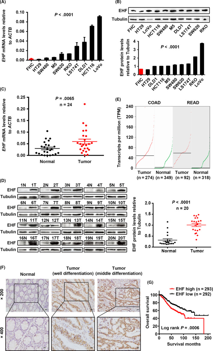
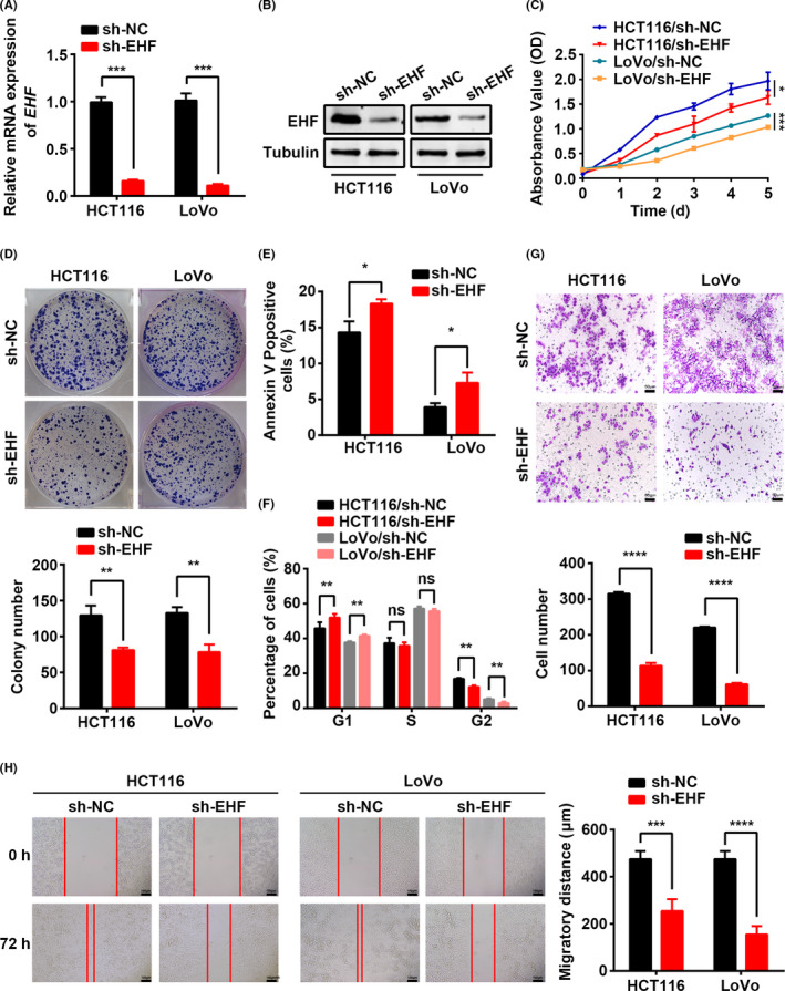
ETS homologous factor (EHF) plays a critical function in epithelial cell differentiation and proliferation. However, the roles of EHF in cancer remain largely unknown. In the present study, we investigated the expression levels, precise function and mechanism of EHF in colorectal carcinoma (CRC). We observed significantly elevated EHF expression in CRC cell lines and tissues. EHF overexpression correlated positively with poor differentiation, advanced T stage, and shorter overall survival of CRC patients. Function experiments revealed that EHF overexpression promoted CRC cell proliferation, migration, and invasion in vitro and in vivo. Mechanistically, EHF could directly upregulate transforming growth factor β1 (TGF-β1) expression at the transcription level, thereby activating canonical TGF-β signaling. Our findings provide novel insights into the mechanisms of EHF in tumorigenesis, invasion, and metastasis of CRC, which may help to provide new therapeutic targets for CRC intervention.

Keywords: EHF; TGF-β signaling; colorectal carcinoma; proliferation and migration; transcription factor.


Conflict of interest statement

The authors have no conflicts of interest.

Figures

Figure 1
Figure 1
ETS homologous factor is upregulated in CRC and positively associated with poor prognosis in CRC patients. A, B, Expression levels of EHF mRNA (A) and protein (B) in CRC and colon mucosa epithelial (FHC) cell lines. C, D, Expression levels of EHF mRNA (C) and protein (D) in paired CRC and adjacent noncancerous tissues. E, Analysis of EHF expression in colon and rectum adenocarcinoma compared with in normal mucosa according to TCGA and GTEx databases. F, Expression analysis of EHF protein in CRC and normal colorectal mucosa tissues by IHC. Scale bars, 50 μm (×200 magnification) or 20 μm (×400 magnification). G, Correlation between EHF expression and overall survival in CRC patients. The median value of EHF expression was chosen as the point for separating tumors into low‐level and high‐level expression groups (log‐rank P = .0006)
Figure 2
Figure 2
EHF depletion mediated by shRNA suppresses CRC cell proliferation, migration, and invasion in vitro. A, B, Decreased expression of EHF after infection of lentivirus vector harboring shRNA‐EHF was confirmed in 2 CRC cell lines by real‐time RT‐PCR (A) and western blotting (B). C, D, Effects of EHF depletion on CRC cell proliferation by CCK‐8 (C) and colony formation assays (D). E, F, The effects of EHF depletion on cell apoptosis (E) and cell cycle progression (F) by flow cytometry. G, Transwell assays were used to determine the effects of EHF depletion on the invasion ability of CRC cells. Representative images (upper) and quantitative analyses (lower) H, The effects of EHF depletion on the migration potencies of CRC cells by using wound‐healing assay. Representative images (left) and quantitative analyses (right) are shown. Data are expressed as the means ± SD in 3 independent experiments. *P < .05, **P < .01, ***P < .001, and ****P < .0001
Figure 3
Figure 3
EHF depletion suppresses CRC cell proliferation and metastasis in vivo. A, EHF downregulation inhibited subcutaneous tumor formation in nude mice. LoVo cells with EHF depletion and control cells were inoculated into nude mice (n = 10 per group). The graphs show the tumor xenografts 30 d after ectopic‐subcutaneous implantation in nude mice with EHF depletion and control cells. B, The gross of xenografts. C, The effect of EHF depletion on CRC tumor growth was evaluated based on tumor volume in the 2 groups. D, Tumor cells were transplanted by intrasplenic injection to evaluate liver metastasis potential. The representative photographs of gross and H&E staining are shown
Figure 4
Figure 4
EHF overexpression promotes cell proliferation, migration, and invasion of CRC cells in vitro. A, B, Increased expression of EHF after transfection of the pcDNA4.0‐EHF vector was confirmed in 2 CRC cell lines by real‐time RT‐PCR (A) and western blotting (B). C, EHF overexpression promoted cell proliferation in HT29 and SW480 cells. D, EHF overexpression increased the capacity to form colonies in HT29 and SW480 cells. E, EHF overexpression repressed the apoptosis ratio relative to control cells. F, G, Matrigel invasion chamber and wound‐healing assays showed that EHF overexpression induced cell invasiveness (F) and migration (G) in both SW480 and HT29 cells. Representative images (left) and quantitative analyses (right) are shown. Data are presented as the mean ± SD. The results were reproducible in 3 independent experiments. * P < .05, ** P < .01, *** P < .001, and ****P < .0001
Figure 5
Figure 5
EHF activates TGF‐β1 transcription. A, The different expressed genes (DEGs) in the GSE21510 microarray dataset (n = 104) analyzed by Qlucore Omics Explorer software. B, DEGs were involved in different pathways and biological processes, as indicated by GO enrichment analysis. C, GSEA showed the enrichment of TGF‐β pathway in CRC cells with EHF upregulation. D, Expression correlation analysis between EHF (x) and TGF‐β1 (y) in 695 CRC tissues (R = 0.1859, P < .0001). E, Expression levels of TGF‐β1 in SW480 cells after EHF overexpression and in LoVo cells after knockdown of EHF. F, Schematic depiction of the TGF‐β1 promoter with 2 putative EHF binding sites (EBS). G, ChIP analysis of EHF occupancy on the TGF‐β1 promoter. H, EMSA assay showed that EHF protein directly interacts with the sequence of the first EBS in TGF‐β1 promoter. I, Dual‐luciferase assays showed that EHF activated TGF‐β1 transcription in 293T cells. The results were reproducible in 3 independent experiments. *P < .05, **P < .01, ***P < .001, and ***P < .0001
Figure 6
Figure 6
EHF activates the activity of canonical TGF‐β signaling. A, Western blotting was performed to detect SMAD3, p‐SMAD3, SMAD2, p‐SMAD2, nuclear SMAD4, and cytoplasmic SMAD4 in EHF‐depleted and control CRC cells. B‐E, EHF overexpression and TGF‐β1 (10 ng/mL, 48 h) significantly promoted CRC cell proliferation (B), colony formation (C), migration (D) and invasion (E). The potential effects of EHF on CRC cells were completely abolished similar to the control cells by TGF‐β inhibitor (SE525334, 1 μm, 48 h, B‐E). F, TGF‐β1 induced EHF expression (upper) and TGF‐β inhibitor repressed EHF expression (lower). G, Schematic diagram showing the mechanism of action of EHF in CRC

References

    1. Bray F, Ferlay J, Soerjomataram I, Siegel RL, Torre LA, Jemal A. Global cancer statistics 2018: GLOBOCAN estimates of incidence and mortality worldwide for 36 cancers in 185 countries. CA Cancer J Clin. 2018;68(6):394‐424. - PubMed
    1. Dienstmann R, Vermeulen L, Guinney J, Kopetz S, Tejpar S, Tabernero J. Consensus molecular subtypes and the evolution of precision medicine in colorectal cancer. Nat Rev Cancer. 2017;17:79‐92. - PubMed
    1. Massague J, Obenauf AC. Metastatic colonization by circulating tumour cells. Nature. 2016;529:298‐306. - PMC - PubMed
    1. Sizemore GM, Pitarresi JR, Balakrishnan S, Ostrowski MC. The ETS family of oncogenic transcription factors in solid tumours. Nat Rev Cancer. 2017;17:337‐351. - PubMed
    1. Luk IY, Reehorst CM, Mariadason JM. ELF3, ELF5, EHF and SPDEF transcription factors in tissue homeostasis and cancer. Molecules. 2018;23:2191. - PMC - PubMed

MeSH terms