

Mitochondrial Homeostasis and Signaling in Parkinson's Disease
- PMID: 32372945
- PMCID: PMC7186467
- DOI: 10.3389/fnagi.2020.00100
Mitochondrial Homeostasis and Signaling in Parkinson's Disease
Abstract
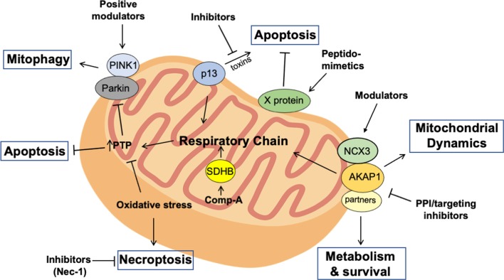
The loss of dopaminergic (DA) neurons in the substantia nigra leads to a progressive, long-term decline of movement and other non-motor deficits. The symptoms of Parkinson's disease (PD) often appear later in the course of the disease, when most of the functional dopaminergic neurons have been lost. The late onset of the disease, the severity of the illness, and its impact on the global health system demand earlier diagnosis and better targeted therapy. PD etiology and pathogenesis are largely unknown. There are mutations in genes that have been linked to PD and, from these complex phenotypes, mitochondrial dysfunction emerged as central in the pathogenesis and evolution of PD. In fact, several PD-associated genes negatively impact on mitochondria physiology, supporting the notion that dysregulation of mitochondrial signaling and homeostasis is pathogenically relevant. Derangement of mitochondrial homeostatic controls can lead to oxidative stress and neuronal cell death. Restoring deranged signaling cascades to and from mitochondria in PD neurons may then represent a viable opportunity to reset energy metabolism and delay the death of dopaminergic neurons. Here, we will highlight the relevance of dysfunctional mitochondrial homeostasis and signaling in PD, the molecular mechanisms involved, and potential therapeutic approaches to restore mitochondrial activities in damaged neurons.
Keywords: AKAP; PKA; Parkinson’s disease; cAMP; mitocondria.
Copyright © 2020 Scorziello, Borzacchiello, Sisalli, Di Martino, Morelli and Feliciello.
Figures
Similar articles
-
Connecting the dots between mitochondrial dysfunction and Parkinson's disorder: focus mitochondria-targeting therapeutic paradigm in mitigating the disease severity.Environ Sci Pollut Res Int. 2021 Jul;28(28):37060-37081. doi: 10.1007/s11356-021-14619-6. Epub 2021 May 29. Environ Sci Pollut Res Int. 2021. PMID: 34053042 Review.
-
Dopaminergic Neurons Exhibit an Age-Dependent Decline in Electrophysiological Parameters in the MitoPark Mouse Model of Parkinson's Disease.J Neurosci. 2016 Apr 6;36(14):4026-37. doi: 10.1523/JNEUROSCI.1395-15.2016. J Neurosci. 2016. PMID: 27053209 Free PMC article.
-
Impaired mitochondrial dynamics and function in the pathogenesis of Parkinson's disease.Exp Neurol. 2009 Aug;218(2):235-46. doi: 10.1016/j.expneurol.2009.03.006. Epub 2009 Mar 18. Exp Neurol. 2009. PMID: 19303005 Review.
-
Mfn2 protects dopaminergic neurons exposed to paraquat both in vitro and in vivo: Implications for idiopathic Parkinson's disease.Biochim Biophys Acta Mol Basis Dis. 2017 Jun;1863(6):1359-1370. doi: 10.1016/j.bbadis.2017.02.016. Epub 2017 Feb 16. Biochim Biophys Acta Mol Basis Dis. 2017. PMID: 28215578 Free PMC article.
-
Fighting Parkinson's disease: The return of the mitochondria.Mitochondrion. 2022 May;64:34-44. doi: 10.1016/j.mito.2022.02.003. Epub 2022 Feb 24. Mitochondrion. 2022. PMID: 35218960 Review.
Cited by
-
Neuroprotective and anti-inflammatory properties of proteins secreted by glial progenitor cells derived from human iPSCs.Front Cell Neurosci. 2024 Aug 6;18:1449063. doi: 10.3389/fncel.2024.1449063. eCollection 2024. Front Cell Neurosci. 2024. PMID: 39165834 Free PMC article.
-
Mitochondrial Dysfunction is a Crucial Immune Checkpoint for Neuroinflammation and Neurodegeneration: mtDAMPs in Focus.Mol Neurobiol. 2025 Jun;62(6):6715-6747. doi: 10.1007/s12035-024-04412-0. Epub 2024 Aug 8. Mol Neurobiol. 2025. PMID: 39115673 Review.
-
Elucidating the Multi-Targeted Role of Nutraceuticals: A Complementary Therapy to Starve Neurodegenerative Diseases.Int J Mol Sci. 2021 Apr 14;22(8):4045. doi: 10.3390/ijms22084045. Int J Mol Sci. 2021. PMID: 33919895 Free PMC article. Review.
-
Dopaminergic Axons: Key Recitalists in Parkinson's Disease.Neurochem Res. 2022 Feb;47(2):234-248. doi: 10.1007/s11064-021-03464-1. Epub 2021 Oct 12. Neurochem Res. 2022. PMID: 34637100 Review.
-
Altered Mitochondrial Bioenergetics and Calcium Kinetics in Young-Onset PLA2G6 Parkinson's Disease iPSCs.J Neurochem. 2025 Apr;169(4):e70059. doi: 10.1111/jnc.70059. J Neurochem. 2025. PMID: 40189860 Free PMC article.
References
LinkOut - more resources
Full Text Sources