. 2020 Apr 21:11:189.
doi: 10.3389/fendo.2020.00189. eCollection 2020.

Mutation of Isocitrate Dehydrogenase 1 in Cholangiocarcinoma Impairs Tumor Progression by Inhibiting Isocitrate Metabolism

Affiliations

Mutation of Isocitrate Dehydrogenase 1 in Cholangiocarcinoma Impairs Tumor Progression by Inhibiting Isocitrate Metabolism

Li Su et al. Front Endocrinol (Lausanne). .

Abstract

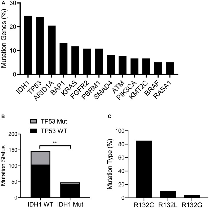
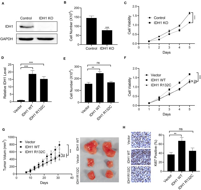
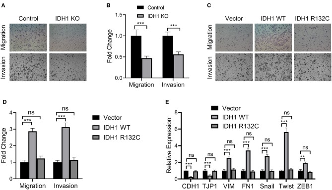
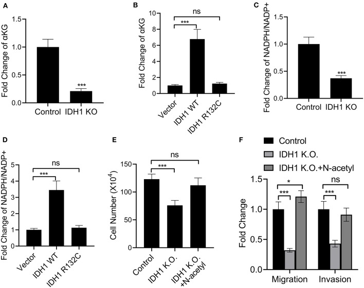
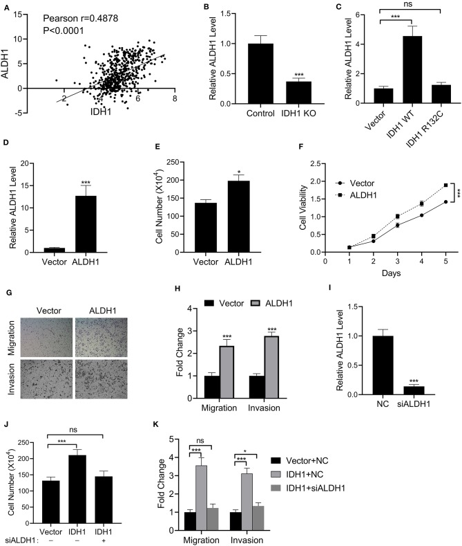
Aim: Isocitrate dehydrogenase 1 (IDH1) is key enzyme involved in cellular metabolism and DNA repair. Mutations in IDH1 occur in up to 25% of cholangiocarcinomas. The present study aimed to explore the features of cellosaurus REB cells with mutant and wide-type IDH1. Methods: To compare the features of IDH1 knockout and mutation in cholangiocarcinoma, we firstly constructed the IDH1 knockout and IDH1 mutation cell lines. We then evaluated the viability of these cell lines using the cell count assay and MTT assay. Next, we determined cell migration and invasion using the Transwell assay. Additionally, to evaluate the effects of IDH1 on cellular metabolism, the levels of α-ketoglutarate (α-KG) and nicotinamide adenine dinucleotide phosphate (NADPH) were determined using enzyme-linked immunosorbent assay. We then applied ChIPbase dataset to explore the genes that were regulated by IDH1. Results: High frequency of mutated IDH1 was observed in the cholangiocarcinoma and IDH1 R132C was presented in more than 80% of mutations. The results showed that IDH1 knockout decreased cell proliferation, migration and invasion, whereas the overexpression of IDH1 in IDH1 knockout cell line recovered its proliferation, migration and invasion capacities. Additionally, IDH1 mutation reduced the levels of NADPH and α-KG. Furthermore, investigation into the underlying mechanisms revealed that IDH1 overexpression induced the expression of aldehyde dehydrogenase 1 thereby promoting cell proliferation, migration and invasion. Conclusion:IDH1 plays an important role in cholangiocarcinoma and its mutation impairs tumor progression in part by inhibition of isocitrate metabolism.

Keywords: NADPH; aldehyde dehydrogenase 1; cholangiocarcinoma; isocitrate dehydrogenase 1; α-ketoglutarate.


Figures

Figure 1
Figure 1
IDH1 was mutated in cholangiocarcinoma. The TCGA cholangiocarcinoma dataset (MSK, Clin Cancer Res 2018) was downloaded and analyzed in cBioportal (www.cbioportal.org). (A) The frequency of mutated genes in Cholangiocarcinoma. (B) The proportion of TP53 mutated patients in IDH1 wildtype (WT) or mutated group. (C) The proportion of IDH1 with different mutation types. The data were represented as mean ± S.D. **P < 0.01.
Figure 2
Figure 2
The overexpression of IDH1 promoted cell proliferation of cholangiocarcinoma. (A) Western blot was used to determine the expressions of IDH1 in the IDH knockout (KO) REB cells. Cell count assay (B) and MTT assay (C) were used to determine the cell viabilities of REB and IDH1 KO REB cells. (D) qPCR was used to determine the relative expressions of IDH1 in IDH1 KO REB cells transfected with vector, plasmids containing wildtype IDH1 or plasmids containing IDH1 R132C mutation sequence. Cell count assay (E) and MTT assay (F) were used to determine cell viabilities of IDH1 KO cells transfected with vector, plasmids containing wildtype IDH1 or plasmids containing IDH1 R132C mutation type. (G) The tumor volume of xenograft mouse transplanted with IDH1 KO REB cells transfected with vector, wildtype IDH1 or IDH1 R132C mutation plasmid. (H) Immunohistochemistry demonstrated percentage of Ki67 positive in tumor tissue from each group. Data are shown as mean ± S.D. **P < 0.01, ***P < 0.001; ns indicates no significance.
Figure 3
Figure 3
IDH1 promoted cell migration and invasion and IDH1 knockout decreased cell migration and proliferation. (A) Cell migration and invasion of the REB and IDH1 KO REB cells were determined using Transwell and (B) the values in each group were shown as mean ± S.D. (C) Cell migration and invasion of the IDH1 KO REB cells transfected with vector, wildtype IDH1, or IDH1 R132C mutation plasmid and (D) the values in each group were shown as mean ± S.D. (E) qPCR was used to analyze the relative expressions of a panel of EMT relevant markers in IDH1 KO REB cells transfected with vector, wildtype IDH1 or IDH1 R132C mutation plasmid. Data were shown as mean ± S.D. **P < 0.01, ***P < 0.001; ns indicates no significance.
Figure 4
Figure 4
IDH1 mutation reduced NADPH and α-KG levels. (A,B) ELISA was used to determine the levels of α-KG in REB cells, IDH1 KO REB cells, IDH1 KO REB cells transfected with vector, wildtype IDH1 or IDH1 R132C mutation plasmid. (C,D) ELISA was used to determine the levels of NADPH in REB cells, IDH1 KO REB cells, IDH1 KO REB cells transfected with vector, wildtype IDH1 or IDH1 R132C mutation plasmid. (E) Cell count assay were used to determine the cell viabilities of REB cells or REB IDH1 K.O. cells treated with or without N-acetyl cysteine. (F) Cell migration and invasion assay of the REB cells or REB IDH1 K.O. cells treated with or without N-acetyl cysteine. Data were shown as mean ± S.D. *P < 0.05, ***P < 0.001; ns indicates no significance.
Figure 5
Figure 5
IDH1 regulated the expressions of ALDH1. (A) The correlation of IDH1 and ALDH1 expression in the patients with cholangiocarcinoma was analyzed using ChIPbase dataset. (B,C) qPCR was used to determine the levels of ALDH1 in REB cells, IDH1 KO REB cells, and IDH1 KO REB cells transfected with vector, wildtype IDH1 or IDH1 R132C mutation plasmid. (D) qPCR was used to determine the relative expressions of ALDH1 in REB cells transfected with ALDH1 expression plasmid. (E,F) Cell count assay and MTT assay were used to determine the cell viabilities of REB cells transfected with vector or ALDH1 expression plasmid. (G,H) Cell migration and invasion assay of REB cells transfected with vector or ALDH1 expression plasmid were determined using Transwell and the values in each group were shown as mean ± S.D. (I) qPCR was used to determine the relative expressions of ALDH1 in REB cells transfected with ALDH1 expression plasmid. (J) Cell count assay were used to determine the cell viabilities of REB cells transfected with vector or IDH1 expression plasmid in the presence or absence of ALDH1 siRNA. (K) Cell migration and invasion assay of the REB cells transfected with vector or IDH1 expression plasmid in the presence or absence of ALDH1 siRNA. *P < 0.05, ***P < 0.001; ns indicates not significance.

Similar articles

Cited by

References

    1. Khan SA, Thomas HC, Davidson BR, Taylor-Robinson SD. Cholangiocarcinoma. Lancet. (2005) 366:1303–14. 10.1016/S0140-6736(05)67530-7 - DOI - PubMed
    1. Blechacz B, Gores GJ. Cholangiocarcinoma: advances in pathogenesis, diagnosis, and treatment. Hepatology. (2008) 48:308–21. 10.1002/hep.22310 - DOI - PMC - PubMed
    1. Razumilava N, Gores GJ. Cholangiocarcinoma. Lancet. (2014) 383:2168–79. 10.1016/S0140-6736(13)61903-0 - DOI - PMC - PubMed
    1. Mazzaferro V, Gorgen A, Roayaie S, Droz Dit Busset M, Sapisochin G. Liver resection and transplantation for intrahepatic cholangiocarcinoma. J Hepatol. (2020) 72:364–77. 10.1016/j.jhep.2019.11.020 - DOI - PubMed
    1. Khan SA, Davidson BR, Goldin RD, Heaton N, Karani J, Pereira SP, et al. . Guidelines for the diagnosis and treatment of cholangiocarcinoma: an update. Gut. (2012) 61:1657–69. 10.1136/gutjnl-2011-301748 - DOI - PubMed

Publication types

MeSH terms