. 2020 Apr 16:11:626.
doi: 10.3389/fimmu.2020.00626. eCollection 2020.

Bacteria-Induced Acute Inflammation Does Not Reduce the Long-Term Reconstitution Capacity of Bone Marrow Hematopoietic Stem Cells

Affiliations

Bacteria-Induced Acute Inflammation Does Not Reduce the Long-Term Reconstitution Capacity of Bone Marrow Hematopoietic Stem Cells

Xiaoyu Zhang et al. Front Immunol. .

Abstract

Pathogen-initiated chronic inflammation or autoimmune diseases accelerate proliferation and promote differentiation of hematopoietic stem cells (HSCs) but simultaneously reduce reconstitution capacity. Nevertheless, the effect of acute infection and inflammation on functional HSCs is still largely unknown. Here we found that acute infection elicited by heat-inactivated Escherichia coli (HIEC) expanded bone marrow lineage-negative (Lin)- stem-cell antigen 1 (Sca-1)+cKit+ (LSK) cell population, leading to reduced frequency of functional HSCs in LSK population. However, the total number of BM phenotypic HSCs (Flk2-CD48-CD150+ LSK cells) was not altered in HIEC-challenged mice. Additionally, the reconstitution capacity of the total BM between infected and uninfected mice was similar by both the competitive repopulation assay and measurement of functional HSCs by limiting dilution. Thus, occasionally occurring acute inflammation, which is critical for host defenses, is unlikely to affect HSC self-renewal and maintenance of long-term reconstitution capacity. During acute bacterial infection and inflammation, the hematopoietic system can replenish hematopoietic cells consumed in the innate inflammatory response by accelerating hematopoietic stem and progenitor cell proliferation, but preserving functional HSCs in the BM.

Keywords: acute infection; hematopoietic stem cells; inflammation; long-term reconstitution; self-renewal.


Figures

Figure 1
Figure 1
HIEC-elicited acute inflammation triggers expansion of lineage–sca-1+c-kit+ (LSK) cells. (A) Experimental scheme for the in vivo HI E. coli challenge experiments. Heat-inactivated E. coli (HIEC, 1 × 10⁷ CFU) were injected intraperitoneally. At each time point, BM cells were collected and analyzed by FACS. (B) Representative FACS plots showing changes in LK and LSK cell frequency in BM mononuclear cells at different time points after HIEC challenge. (C) Percentage of LSK and LK cells in BM mononuclear cell (BMMC) populations at different time points after E. coli challenge. Data shown are means ± SD (n = 4 mice). *p < 0.05, **p < 0.01, ***p < 0.001, ****p < 0.0001 vs. unchallenged mice (blk). (D) Absolute number of LSK and LK cells in the total BM at different time points after E. coli challenge. Data shown are means ± SD (n = 4 mice). *p < 0.05, **p < 0.01, ***p < 0.001, ****p < 0.0001 vs. unchallenged mice (blk). (E) Percentage of G1 and G2/S/M cells in LK and LSK cells 24 h after the HI E. coli challenge. Data shown are means ± SD (n = 5 mice). *p < 0.05, **p < 0.01, ***p < 0.001 vs. unchallenged mice (time 0). (F) The number of committed myeloid progenitors analyzed using a quantitative granulocyte–monocyte colony-forming unit (CFU-GM) assay. BMMCs were prepared 24 h after E. coli injection and cultured in MethoCult™ GF M3434 medium for 7 days. Representative photographs of cell clusters/colonies are shown. The number of granulocyte colony-forming units (CFU-G), monocyte colony-forming units (CFU-M), and granulocyte–monocyte colony-forming units (CFU-GM) from 10,000 BMMCs were calculated. Data shown are mean ± SD of n = 3 mice. *p < 0.01 vs. PBS-treated mice. (G) Representative FACS plots showing HSC/HPC subpopulations. LT-HSC (CD135CD150+CD48LSK), ST-HSC (CD135CD150CD48LSK), MPP2 (CD135CD150+CD48+LSK, myeloid-biased), MPP3 (CD135CD150CD48+LSK, myeloid-biased), and MPP4 (CD135+CD150CD48+LSK, lymphoid-primed) cell populations were defined as previously reported (54). (H) Percentage of LT-HSC, ST-HSC, MPP2, MPP3, and MPP4 cells in BM mononuclear cell (BMMC) population and their absolute number in tibia 24 h after the HI E. coli challenge. Data shown are means ± SD (n = 5–7 mice). *p < 0.05, **p < 0.01, ***p < 0.001, ****p < 0.0001 vs. control (PBS treated).
Figure 2
Figure 2
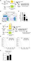
HIEC-elicited acute inflammation reduces the frequency of functional HSCs in expanded LSK cells but does not alter the total number of functional HSCs in the BM. (A) Schematic of the in vivo hematopoiesis reconstitution experiment. 1000 LSK cells from PBS or HIEC-challenged mice (CD45.1) were transplanted into lethally-irradiated congenic recipients (CD45.2) together with 0.5 million supporting cells (CD45.2). BM chimerism in recipients was analyzed at each indicated time point. (B) Representative FACS plots of donor-derived WBCs in the peripheral blood 6 months after transplantation. (C) The percentages of donor derived cells (CD45.1+) in total hematopoietic cells (CD45.2+ or CD45.1+) at the indicated time points in PBS and HIEC-treated groups. Data shown are means ± SD (n = 5 mice). **p < 0.01 vs. control by unpaired two-tailed nonparametric t-test. (D) Schematic of the limiting dilution analysis (LDA). Limiting dilution experiments were performed with indicated doses of LSK cells obtained from PBS- or HIEC-treated mice (CD45.1) combined with 5 × 10⁵ competing cells (CD45.2) transplanted into recipients (CD45.2). BM chimerism in recipients was analyzed at each indicated time point. Hematopoietic chimerism was analyzed by FACS. (E) Log-fraction plots of the limiting dilution model fitted to the data in Figure S2B and Table S1. Recipient chimerism was determined at 6 months, and >0.5% engraftment was considered positive. The slope of the line is the log-positive transplantation fraction. The dotted lines give the 95% confidence intervals. The frequencies of functional HSCs in LSK cells were calculated accordingly. All the analyses and calculations were performed using ELDA software. (F) The number of functional HSCs in the BM was assessed using limiting dilution analysis. The number of functional HSCs was calculated based on the raw data shown in Table S1. Data shown are mean ± SD of n = 4 mice. NSp > 0.05 vs. PBS-treated mice.
Figure 3
Figure 3
HIEC-induced acute inflammation does not affect the long-term reconstitution activity of BM HSCs in primary recipients. (A) Schematic of the competitive BM transplantation experiment. 0.5 million total BM cells from PBS-treated or HIEC-challenged mice (CD45.2) were transplanted into lethally-irradiated congenic recipients (CD45.1 and 2 C57/B6 mice) together with 0.5 million competing BM cells (CD45.1). BM chimerism in recipients was analyzed at each indicated time point. Hematopoietic chimerism was analyzed by FACS. (B) Representative FACS plots showing the percentage of donor-derived WBCs in the peripheral blood 24 weeks after transplantation in PBS and HIEC groups. The experiments were conducted 24 weeks after the transplantation. (C) The percentage of the donor-derived CD45.2+ and CD45.1+ WBCs in the peripheral blood at the indicated time points. Data shown are mean ± SD of n = 5 mice. (D) Ratio of CD45.2+ to CD45.1+ cells at the indicated time points after transplantation in PBS and HIEC groups. Data shown are mean ± SD of n = 5 mice. (E) Chimerism in indicated blood lineages. Ratio of CD45.2+ to CD45.1+ cells was calculated based on the percentage of donor-derived CD45.2+ and CD45.1+ cells in each indicated linage (Figure S3). Data shown are mean ± SD of n = 5 mice. *p < 0.05 vs. PBS-treated group.
Figure 4
Figure 4
HIEC-induced acute inflammation does not affect the long-term reconstitution activity of BM HSCs after the secondary BM transplantation. (A) Schematic of secondary BM transplantation experiment. One million total BM cells from primary recipients were transplanted into lethally-irradiated congenic recipients (CD45.1 and 2 C57/B6 mice). BM chimerism in secondary recipients was analyzed at each indicated time point. Peripheral blood was obtained retro-orbitally using a heparinized capillary tube. Hematopoietic chimerism was analyzed by FACS. (B) Representative FACS plots showing the percentage of CD45.2+ and CD45.1+ WBCs in the peripheral blood 20 weeks after secondary BM transplantation. The experiments were conducted 20 weeks after the transplantation. (C) The percentage of donor-derived CD45.2+ and CD45.1+ WBCs in the PB of secondary recipients at the indicated time points. Data shown are mean ± SD of n = 5 mice. (D) Ratio of CD45.2+ to CD45.1+ cells at the indicated time points after secondary BM transplantation. Data shown are mean ± SD of n = 5 mice. (E) Chimerism in indicated blood lineages. Ratio of CD45.2+ to CD45.1+ cells was calculated based on the percentage of donor-derived CD45.2+ and CD45.1+ cells in each indicated linage (Figure S4). Data shown are mean ± SD of n = 5 mice. *p < 0.05 vs. PBS-treated group.

References

    1. Kondo M, Wagers AJ, Manz MG, Prohaska SS, Scherer DC, Beilhack GF, et al. . Biology of hematopoietic stem cells and progenitors: implications for clinical application. Annu Rev Immunol. (2003) 21:759–806. 10.1146/annurev.immunol.21.120601.141007 - DOI - PubMed
    1. Glatman Zaretsky A, Engiles JB, Hunter CA. Infection-induced changes in hematopoiesis. J Immunol. (2014) 192:27–33. 10.4049/jimmunol.1302061 - DOI - PMC - PubMed
    1. King KY, Goodell MA. Inflammatory modulation of HSCs: viewing the HSC as a foundation for the immune response. Nat Rev Immunol. (2011) 11:685–92. 10.1038/nri3062 - DOI - PMC - PubMed
    1. Shahbazian LM, Quinton LJ, Bagby GJ, Nelson S, Wang G, Zhang P. Escherichia coli pneumonia enhances granulopoiesis and the mobilization of myeloid progenitor cells into the systemic circulation. Crit Care Med. (2004) 32:1740–6. 10.1097/01.CCM.0000132900.84627.90 - DOI - PubMed
    1. Singh P, Yao Y, Weliver A, Broxmeyer HE, Hong SC, Chang CH. Vaccinia virus infection modulates the hematopoietic cell compartments in the bone marrow. Stem Cells. (2008) 26:1009–16. 10.1634/stemcells.2007-0461 - DOI - PMC - PubMed

Publication types