. 2020 Apr 27;10(12):5623-5640.
doi: 10.7150/thno.44836. eCollection 2020.

Branched chain amino acids exacerbate myocardial ischemia/reperfusion vulnerability via enhancing GCN2/ATF6/PPAR-α pathway-dependent fatty acid oxidation

Affiliations

Branched chain amino acids exacerbate myocardial ischemia/reperfusion vulnerability via enhancing GCN2/ATF6/PPAR-α pathway-dependent fatty acid oxidation

Yueyang Li et al. Theranostics. .

Abstract

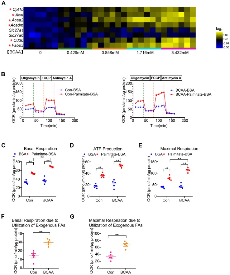
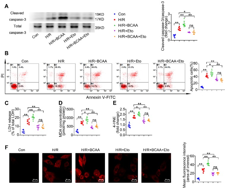
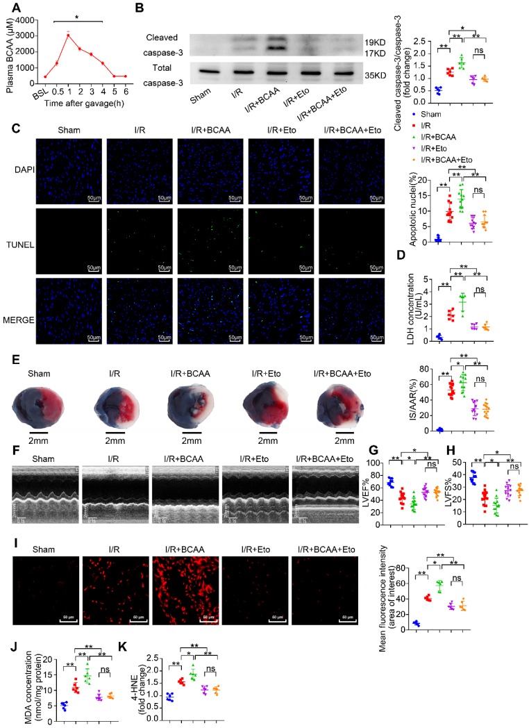
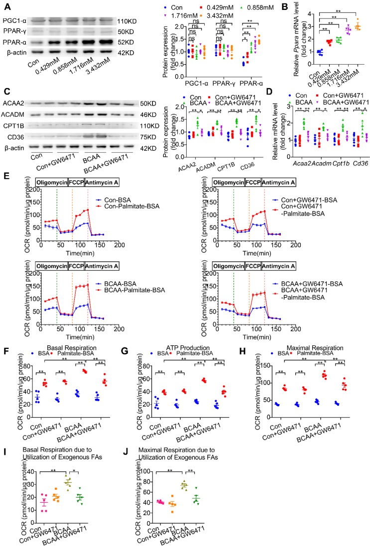
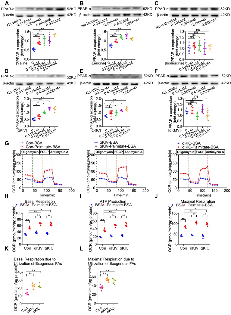
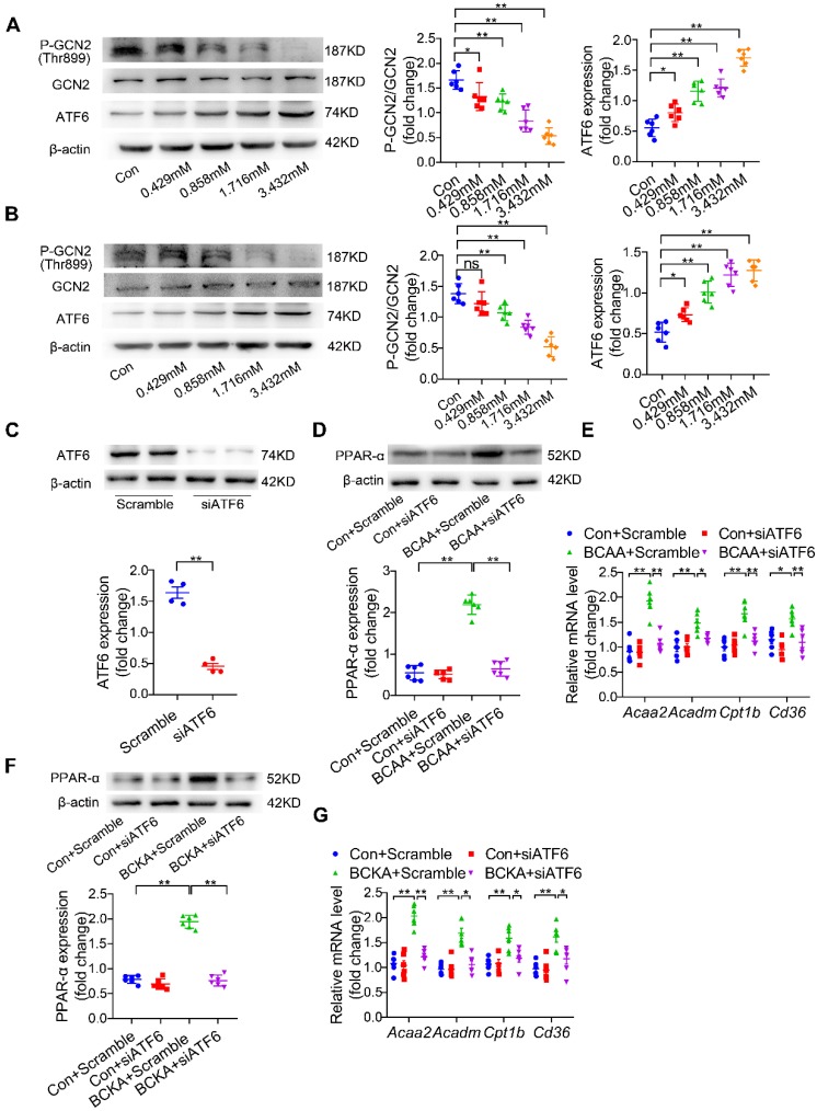
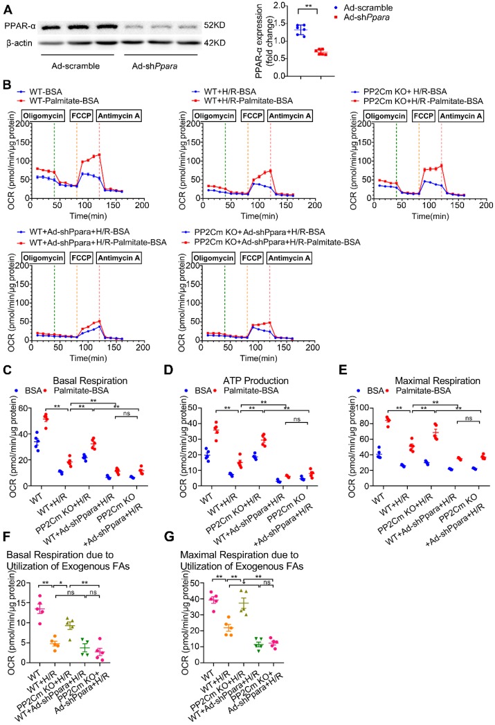
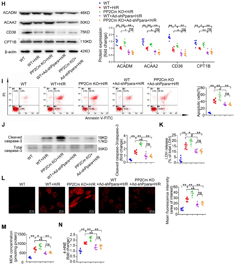
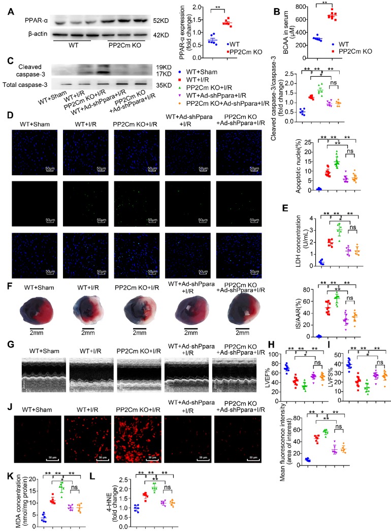
Rationale: Myocardial vulnerability to ischemia/reperfusion (I/R) injury is strictly regulated by energy substrate metabolism. Branched chain amino acids (BCAA), consisting of valine, leucine and isoleucine, are a group of essential amino acids that are highly oxidized in the heart. Elevated levels of BCAA have been implicated in the development of cardiovascular diseases; however, the role of BCAA in I/R process is not fully understood. The present study aims to determine how BCAA influence myocardial energy substrate metabolism and to further clarify the pathophysiological significance during cardiac I/R injury. Methods: Parameters of glucose and fatty acid metabolism were measured by seahorse metabolic flux analyzer in adult mouse cardiac myocytes with or without BCAA incubation. Chronic accumulation of BCAA was induced in mice receiving oral BCAA administration. A genetic mouse model with defective BCAA catabolism was also utilized. Mice were subjected to MI/R and the injury was assessed extensively at the whole-heart, cardiomyocyte, and molecular levels. Results: We confirmed that chronic accumulation of BCAA enhanced glycolysis and fatty acid oxidation (FAO) but suppressed glucose oxidation in adult mouse ventricular cardiomyocytes. Oral gavage of BCAA enhanced FAO in cardiac tissues, exacerbated lipid peroxidation toxicity and worsened myocardial vulnerability to I/R injury. Etomoxir, a specific inhibitor of FAO, rescued the deleterious effects of BCAA on I/R injury. Mechanistically, valine, leucine and their corresponding branched chain α-keto acid (BCKA) derivatives, but not isoleucine and its BCKA derivative, transcriptionally upregulated peroxisome proliferation-activated receptor alpha (PPAR-α). BCAA/BCKA induced PPAR-α upregulation through the general control nonderepresible-2 (GCN2)/ activating transcription factor-6 (ATF6) pathway. Finally, in a genetic mouse model with BCAA catabolic defects, chronic accumulation of BCAA increased FAO in myocardial tissues and sensitized the heart to I/R injury, which could be reversed by adenovirus-mediated PPAR-α silencing. Conclusions: We identify BCAA as an important nutrition regulator of myocardial fatty acid metabolism through transcriptional upregulation of PPAR-α. Chronic accumulation of BCAA, caused by either dietary or genetic factors, renders the heart vulnerable to I/R injury via exacerbating lipid peroxidation toxicity. These data support the notion that BCAA lowering methods might be potentially effective cardioprotective strategies, especially among patients with diseases characterized by elevated levels of BCAA, such as obesity and diabetes.

Keywords: Branched chain amino acids; Fatty acid metabolism; Ischemia/reperfusion injury; Peroxisome proliferation-activated receptor-α; Vulnerability..


Conflict of interest statement

Competing Interests: The authors have declared that no competing interest exists.

Figures

Figure 1
Figure 1
BCAA promote FAO in cardiac myocytes. (A) Adult mouse ventricular myocytes were isolated and treated with different concentrations of BCAA (0, 0.429 mM, 0.858 mM, 1.716 mM, 3.432 mM) for 12 h. Expression of Cpt1b, Acsl, Acaa2, Acadm, Slc27a1, Slc27a6, Cd36, Fabp3 mRNA by real-time PCR, normalized to β-actin (n=6). (B-G) Adult mouse cardiac myocytes were treated with or without BCAA (3.432 mM) for 12 h. FAO levels were determined by seahorse analyzer (n=4-5). (B) OCR curve of Con (No BCAA) group and BCAA group were determined. (C) Basal respiration (D) ATP production (E) maximal respiration (F) basal respiration due to exogenous palmitate-BSA and (G) maximal respiration due to exogenous palmitate-BSA were calculated according to instruction. (C-E) Data were analyzed by one-way ANOVA, followed by a Bonferroni post-hoc test. (F-G) Data were analyzed by Student's t test (two-tailed). * P<0.05, ** P<0.01. All values are presented as mean ± SEM.
Figure 2
Figure 2
BCAA exacerbate H/R injury via enhancing FAO. NRVMs were isolated and subjected to 9 h/3 h H/R, with or without BCAA (3.432 mM) and Eto (10 nmol/L). (A) Cleavage of caspase-3 and total caspase-3 were determined by western blotting (n=6). (B) Apoptosis was analyzed by Annexin V-FITC flow cytometry (n=6). (C-E) LDH release, MDA and 4-HNE were determined as methods described (n=6). (F) Superoxide generation was assessed by DHE staining (n=6). Data were analyzed by one-way ANOVA, followed by a Bonferroni post-hoc test. * P<0.05, ** P<0.01.
Figure 3
Figure 3
BCAA worsen I/R injury, which can be rescued by inhibiting FAO. (A) Serum BCAA concentrations at different time points after gavage (weight ratio, leucine: valine: isoleucine=2:1:1, 1.5 mg/g/day, n=6-8). (B-K) Vehicle or BCAA-supplemented (1.5 mg/g/day, 7 days) mice were treated with or without Eto (20 mg/kg body weight, i.p. injection 15 min before I/R surgery) under basal or I/R conditions. (B) Cardiac cleaved and non-cleaved caspase-3 by western blotting (n=6). (C) Representative cardiac apoptosis determined by TUNEL staining. Green fluorescence indicated TUNEL-positive cardiomyocyte nuclei; blue fluorescence showed total cardiomyocytes nuclei (n=10-15). Scale bar: 50 μm. (D) Cardiac apoptosis by LDH release assay (n=6). (E) Infarct area of heart tissue by Evans blue and tetrazolium chloride (TTC). The blue area represented unaffected heart tissue; white area showed infarcted tissue; red pus white area indicated tissue at risk (n=10-15). Scale bar: 2 mm. (F) Representative M-Mode echocardiographic images. (G and H) Echocardiographic assessment of LV ejection fraction and LV fractional shortening (n=10-15). (I) Superoxide production detected by DHE staining (n=6). Scale bar: 50 μm. (J and K) Lipid peroxidation determined by MDA and 4-HNE contents (n=6). (A) Data were analyzed by Student's t test. (B-K) Data were analyzed by one-way ANOVA, followed by a Bonferroni post-hoc test. * P<0.05, ** P<0.01. All values are presented as mean ± SEM.
Figure 4
Figure 4
BCAA upregulate PPAR-α and PPAR-α targeted genes. (A to B) Adult mouse cardiac myocytes were treated with different concentrations of BCAA (0, 0.429 mM, 0.858 mM, 1.716 mM, 3.432 mM) for 12 h (n=6). (A) Expression of PGC1-α, PPAR-γ, PPAR-α in cardiomyocytes by western blotting. (B) Expression of Ppara in cardiomyocytes by real-time PCR. (C-J) Adult cardiac myocytes were treated with Vehicle (Con), GW6471, BCAA, BCAA+GW6471 for 12 h. (C and D) Expression of ACAA2, ACADM, CD36, CPT1B in cardiomyocytes by western blotting and real-time PCR (n=6). (E) OCR curve of adult cardiac myocytes treated with Vehicle (Con), GW6471, BCAA, BCAA+GW6471 were determined (n=5). (F) Basal respiration (G) ATP production (H) maximal respiration (I) basal respiration due to exogenous palmitate-BSA and (J) maximal respiration due to exogenous palmitate-BSA were calculated according to instruction. N=5 per group. * P<0.05, ** P<0.01. Data were analyzed by one-way ANOVA, followed by a Bonferroni post-hoc test. All values are presented as mean ± SEM.
Figure 5
Figure 5
The effects of BCAA/BCKA on FAO level and PPAR-α expression in cardiac myocytes. (A) Expression of PPAR-α in the presence of increasing concentrations of valine (0, 0.117 mM, 0.234 mM, 0.468 mM, 0.936 mM) by western blotting (n=5-6). (B) Expression of PPAR-α at different concentrations of leucine (0, 0.208 mM, 0.416 mM, 0.832 mM, 1.664 mM) (n=5-6). (C) PPAR-α expression at different concentrations of isoleucine (0, 0.104 mM, 0.208 mM, 0.416 mM, 0.832 mM) (n=5-6). (D) Expression of PPAR-α in the presence of increasing concentrations of a-ketoisovaleric acid (αKIV) (0, 0.117 mM, 0.234 mM, 0.468 mM, 0.936 mM) (n=5-6). (E) Expression of PPAR-α at increasing concentrations of α-ketoisocaproic acid (αKIC) (0, 0.208 mM, 0.416 mM, 0.832 mM, 1.664 mM) (n=5-6). (F) PPAR-α expression at increasing concentrations of α-keto-β-methylvaleric acid (αKMV) (0, 0.104 mM, 0.208 mM, 0.416 mM, 0.832 mM) (n=5-6). (G to L) Adult mouse cardiac myocytes were treated with vehicle (Con), αKIV (0.936 mM) and αKIC (1.664 mM) for 12 h. FAO levels were determined by seahorse analyzer (n=5). (G) OCR curve of Con group, αKIV group and αKIC group were determined. (H) Basal respiration (I) ATP production (J) maximal respiration (K) basal respiration due to exogenous FAs and (L) maximal respiration due to exogenous FAs were calculated according to instruction. Data were analyzed by one-way ANOVA, followed by a Bonferroni post-hoc test. * P<0.05. ** P<0.01. All values are presented as mean ± SEM.
Figure 6
Figure 6
BCAA increase PPAR-α expression in a GCN2/ATF6 pathway-dependent manner. (A) Expression of p-GCN2, GCN2 and ATF6 in the presence of increasing concentrations of BCAA (0, 0.429 mM, 0.858 mM, 1.716 mM, 3.432 mM) by western blotting (n=6). (B) Expression of p-GCN2, GCN2 and ATF6 in the presence of increasing concentrations of BCKA (0, 0.429 mM, 0.858 mM, 1.716 mM, 3.432 mM) by western blotting (n=6). BCKA mixture is composed of αKIC, αKIV and αKMV (weight ratio, αKIC: αKIV: αKMV= 2:1:1). (C) NRVMs were treated with control siRNA and ATF6 siRNA. 48 h after transfection, expression of ATF6 was determined by western blotting (n=4). (D-E) ATF6 siRNA transferred NRVMs were treated with or without BCAA (3.432 mM) (n=6). (D) PPAR-α expression was determined by western blotting. (E) Expression of Acaa2, Acadm, Cd36 and Cpt1b by real-time PCR. (F-G) ATF6 siRNA transferred NRVMs were treated with or without BCKA (3.432 mM) (n=6). (F) PPAR-α expression was determined by western blotting. (G) Expression of Acaa2, Acadm, Cd36 and Cpt1b by real-time PCR. (C) Data were analyzed by Student's t test. (A-B) and (D-G) Data were analyzed by one-way ANOVA, followed by a Bonferroni post-hoc test. * P<0.05, ** P<0.01. All values are presented as mean ± SEM.
Figure 7
Figure 7
PPAR-α knockdown suppresses myocardial FAO levels and protects PP2Cm KO cardiomyocytes against injury following H/R. (A) Expression of PPAR-α by western blotting in cardiomyocytes with or without shPpara adenovirus infection (n=6). (B to N) Ventricular myocytes isolated from WT mice or PP2Cm KO mice were infected with scrambled or shPpara adenovirus for 48 h with or without H/R injury. (B to G) FAO levels were determined by seahorse analyzer (n=4-5). (B) OCR curve treated as mentioned above were determined. (C) Basal respiration (D) ATP production (E) maximal respiration (F) basal respiration due to exogenous FAs and (G) maximal respiration due to exogenous FAs were calculated according to instruction. (H) Expression of ACAA2, ACADM, CD36, CPT1B in cardiomyocytes by western blotting (n=6). (I) Annexin V and propidium iodide (PI) staining by flow cytometry for cardiomyocyte apoptosis determination (n=6). (J) Cleaved and non-cleaved caspase-3 by western blotting (n=6). (K) Cell death assessed by LDH release (n=6). (L) Superoxide production detected by DHE staining (n=6). Scale bar: 20 μm. (M and N) Lipid peroxidation determined by MDA and 4-HNE contents (n=6). Data were analyzed by one-way ANOVA, followed by a Bonferroni post-hoc test. * P<0.05, ** P<0.01. All values are presented as mean ± SEM.
Figure 7
Figure 7
PPAR-α knockdown suppresses myocardial FAO levels and protects PP2Cm KO cardiomyocytes against injury following H/R. (A) Expression of PPAR-α by western blotting in cardiomyocytes with or without shPpara adenovirus infection (n=6). (B to N) Ventricular myocytes isolated from WT mice or PP2Cm KO mice were infected with scrambled or shPpara adenovirus for 48 h with or without H/R injury. (B to G) FAO levels were determined by seahorse analyzer (n=4-5). (B) OCR curve treated as mentioned above were determined. (C) Basal respiration (D) ATP production (E) maximal respiration (F) basal respiration due to exogenous FAs and (G) maximal respiration due to exogenous FAs were calculated according to instruction. (H) Expression of ACAA2, ACADM, CD36, CPT1B in cardiomyocytes by western blotting (n=6). (I) Annexin V and propidium iodide (PI) staining by flow cytometry for cardiomyocyte apoptosis determination (n=6). (J) Cleaved and non-cleaved caspase-3 by western blotting (n=6). (K) Cell death assessed by LDH release (n=6). (L) Superoxide production detected by DHE staining (n=6). Scale bar: 20 μm. (M and N) Lipid peroxidation determined by MDA and 4-HNE contents (n=6). Data were analyzed by one-way ANOVA, followed by a Bonferroni post-hoc test. * P<0.05, ** P<0.01. All values are presented as mean ± SEM.
Figure 8
Figure 8
Exacerbated I/R injury in the PP2Cm KO heart is rescued by PPAR-α knockdown. (A) Expression of PPAR-α were determined by western blotting in WT mice and PP2Cm KO mice (n=6). (B) WT and PP2Cm KO serum BCAA were determined (n=6-8). (C to L) WT mice or PP2Cm KO mice were received intra-myocardial injection with scrambled or shPpara adenovirus 7 days before sham or I/R operation. (C) Cleaved and non-cleaved caspase-3 by western blotting (n=6). (D) Cardiac apoptosis determined by TUNEL staining (n=10-15). Scale bar: 50 μm. (E) Cardiac death by LDH release assay (n=6). (F) Infarct area of heart tissue by Evans blue and TTC (n=10-15). Scale bar: 2 mm. (G) Representative M-Mode echocardiographic images. (H and I) Echocardiographic assessment of LV ejection fraction and LV fractional shortening (n=10-15). (J) Superoxide production detected by DHE staining (n=6). Scale bar: 50 μm. (K and L) Lipid peroxidation determined by MDA and 4-HNE (n=6). (A-B) Data were analyzed by Student's t test. (C-L) Data were analyzed by one-way ANOVA, followed by a Bonferroni post-hoc test. * P<0.05, ** P<0.01. All values are presented as mean ± SEM.

References

    1. Moran AE, Forouzanfar MH, Roth GA, Mensah GA, Ezzati M, Murray CJ. et al. Temporal trends in ischemic heart disease mortality in 21 world regions, 1980 to 2010: the global burden of disease 2010 study. Circulation. 2014;129:1483–92. - PMC - PubMed
    1. Ziegler M, Wang X, Lim B, Leitner E, Klingberg F, Ching V. et al. Platelet-targeted delivery of peripheral blood mononuclear cells to the ischemic heart restores cardiac function after ischemia-reperfusion injury. Theranostics. 2017;7:3192–206. - PMC - PubMed
    1. Kawaguchi M, Takahashi M, Hata T, Kashima Y, Usui F, Morimoto H. et al. Inflammasome activation of cardiac fibroblasts is essential for myocardial ischemia/reperfusion injury. Circulation. 2011;123:594–604. - PubMed
    1. Stanley WC, Recchia FA, Lopaschuk GD. Myocardial substrate metabolism in the normal and failing heart. Physiol Rev. 2005;85:1093–129. - PubMed
    1. Dyck JR, Hopkins TA, Bonnet S, Michelakis ED, Young ME, Watanabe M. et al. Absence of malonyl coenzyme A decarboxylase in mice increases cardiac glucose oxidation and protects the heart from ischemic injury. Circulation. 2006;114:1721–8. - PubMed

Publication types

MeSH terms