

Bilateral, fused pelvic, ectopic, laterally rotated kidneys: A case report
- PMID: 32373249
- PMCID: PMC7191258
- DOI: 10.1016/j.radcr.2020.04.012
Bilateral, fused pelvic, ectopic, laterally rotated kidneys: A case report
Abstract
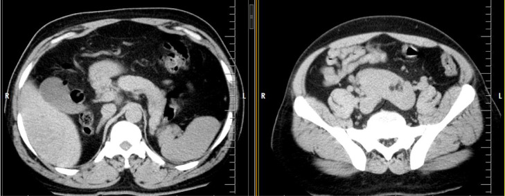
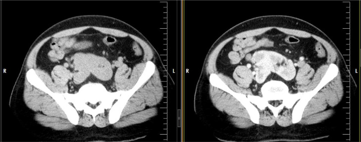
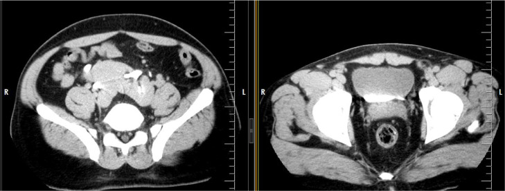
Bilateral fused ectopic pelvic kidneys with lateral rotation abnormality in both kidneys is a fortuitous developmental renal anomaly that to the best of our knowledge this is the first report of this rare anomaly in English literature. Due to the morphologic appearance of this anomaly it is likely it could contribute to the individual remaining asymptomatic therefore discovered incidentally. We present a 42-year-old male who presented with acute unilateral flank pain and incidentally discovered this rare renal anomaly which is an important diagnosis for management by both radiologists and urologists.
Keywords: Ectopic; Fused; Kidneys; Laterally rotated; Pelvic.
© 2020 The Authors. Published by Elsevier Inc. on behalf of University of Washington.
Figures


References
-
- Arrieta MU, Trapote RA, Lizarraga DA. Renal position and fusion anomalies. Ann Pediatr(Barc) 2011;75:329–333. - PubMed
-
- Diaz G. Renal ectopy; report of a case of crossed ectopy without fusion, with fixation of the kidney in normal position by the extraperitoneal route. J Int Coll Surg. 1953;19:158–169. - PubMed
-
- Cinman NM, Okeke Z, Smith AD. Pelvic kidney: associated diseases and treatment. J Endourol. 2007;21(8):836–842. - PubMed
-
- Zafar FS, Lingeman JE.Value of laparoscopy in the management of calculi complicating renal malformations - PubMed
-
- Benz-Bohm G. Pediatric uroradiology. Springer; 2008. Anomalies of kidney rotation, position and fusion; pp. 81–87. ISBN 3540330046.
Publication types
LinkOut - more resources
Full Text Sources
