

doi: 10.1038/s41421-020-0169-8.
eCollection 2020.
The anti-influenza virus drug, arbidol is an efficient inhibitor of SARS-CoV-2 in vitro
Affiliations
- PMID: 32373347
- PMCID: PMC7195821
- DOI: 10.1038/s41421-020-0169-8
Item in Clipboard
The anti-influenza virus drug, arbidol is an efficient inhibitor of SARS-CoV-2 in vitro
Cell Discov.
.
No abstract available
Keywords: Cell biology; Molecular biology.
Conflict of interest statement
Conflict of interestThe authors declare that they have no conflict of interest.
Figures
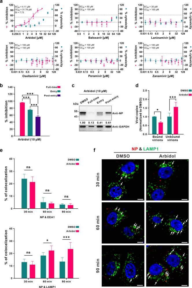
a Antiviral activities of the drugs. The antiviral efficacy was evaluated in Vero E6 cells by qRT-PCR analysis of virus yield at 48 h p.i. Data represent the mean ± standard deviation (SD) from two independent repeats. b, c Time-of-addition experiment of arbidol. Three experimental groups (Full-time, Entry, and Post-entry) were set up as described in the Supplementary Methods. At 16 h p.i., virus yield in the cell supernatant was quantified by qRT-PCR (b), and the expression of NP in infected cells was analyzed by western blots (c). The values below the blot represent the relative band intensity (NP/GAPDH) normalized to that of the DMSO group. d Impact of arbidol on SARS-CoV-2 binding. Vero E6 cells were treated with arbidol (10 μM) or DMSO for 1 h prior to infection with SARS-CoV-2 at 4 °C for 1 h. The supernatant (unbound virions) and the cells containing bound virions (bound virions) were collected for quantification of viral RNA copies by qRT-PCR. e, f Effect of arbidol on intracellular trafficking of SARS-CoV-2. The co-localization of virions with EEs or LEs was analyzed by immunofluorescence assays as described in the Supplementary Methods. e The portion of virions that co-localized with EEs or ELs in each group (n > 150 cells) was quantified by Image J. f Representative confocal microscopic images of virions (red) and LAMP1+ ELs (green) in each group. The nuclei (blue) were stained with Hoechst 33258 dye. White arrows: virions co-localized with ELs; bars: 10 μm. For (b) and (e), statistical analysis was performed using a one-way analysis of variance (ANOVA) with GraphPad Prism. For (d), statistical analysis was performed and calculated by unpaired two-tailed t test. *P < 0.05; ***P < 0.001; ns, not significant.
References
Publication types
LinkOut - more resources
Full Text Sources
Other Literature Sources
Miscellaneous
