. 2020 Apr 25:2020:6151258.
doi: 10.1155/2020/6151258. eCollection 2020.

Neuromyelitis Optica Spectrum Disorder Attack Triggered by Herpes Zoster Infection

Affiliations

Neuromyelitis Optica Spectrum Disorder Attack Triggered by Herpes Zoster Infection

Emanuela Claudia Turco et al. Mult Scler Int. .

Abstract

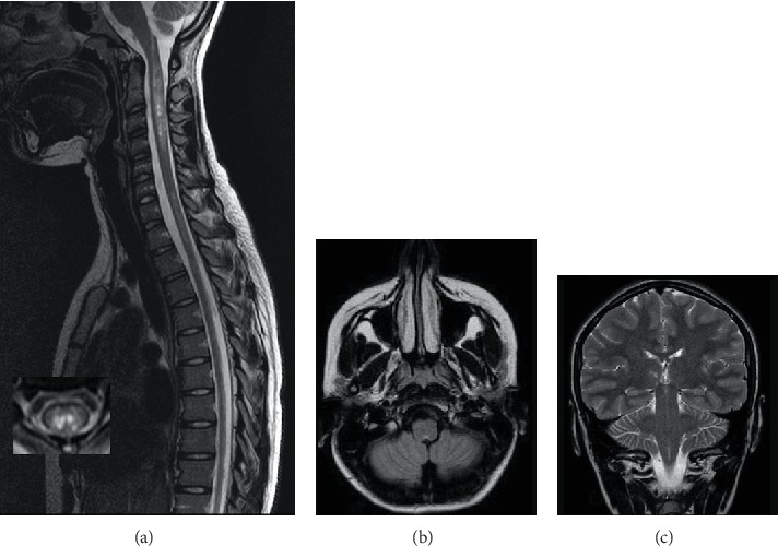
Neuromyelitis optica spectrum disorder is a severe autoimmune disease of the central nervous system characterized by recurrent inflammatory events primarily involving the optic nerves and spinal cord. Recently, a triggering role of infectious events in the development of NMOSD has been suggested. Varicella zoster virus is the most common viral infection involved, though the linkage with anti-aquaporin-4 antibodies is so far unknown. We report, to the best of our knowledge, the first pediatric case report about NMOSD relapse triggered by herpes zoster infection. The strict temporal relationship between VZV infection and NMO attacks seems to be more than simply due to chance; however, further reports are needed to be confirmed.


Conflict of interest statement

The authors declare that there is no conflict of interest.

Figures

Figure 1
Figure 1
(a) Sagittal and axial T2-weighted spinal MR imaging showing hyperintense lesions longitudinally extended from C1 to C5 and from C6 to T6, involving the central spinal cord, with bright spotty lesions. (b) Axial T2-weighted fluid-attenuated inversion recovery (FLAIR) with hyperintense lesion in the brainstem involving the dorsal medulla (area postrema). (c) Coronal T1-weighted brain MRI shows a lesion on the right ventrothalamic area.

References

    1. Tan C. T., Mao Z., Qiu W., Hu X., Wingerchuk D. M., Weinshenker B. G. International consensus diagnostic criteria for neuromyelitis optica spectrum disorders. Neurology. 2016;86(5):491–492. doi: 10.1212/WNL.0000000000002366. - DOI - PubMed
    1. Sellner J., Hemmer B., Mühlau M. The clinical spectrum and immunobiology of parainfectious neuromyelitis optica (Devic) syndromes. Journal of Autoimmunity. 2010;34(4, article S0896841109001279):371–379. doi: 10.1016/j.jaut.2009.09.013. - DOI - PubMed
    1. Pekcevik Y., Orman G., Lee I. H., Mealy M. A., Levy M., Izbudak I. What do we know about brain contrast enhancement patterns in neuromyelitis optica? Clinical Imaging. 2016;40(3):573–580. doi: 10.1016/j.clinimag.2015.07.027. - DOI - PMC - PubMed
    1. Tenembaum S., Chitnis T., Nakashima I., et al. Neuromyelitis optica spectrum disorders in children and adolescents. Neurology. 2016;87(9) Supplement 2:S59–S66. doi: 10.1212/WNL.0000000000002824. - DOI - PubMed
    1. Zhong X., Zhou Y., Lu T., et al. Infections in neuromyelitis optica spectrum disorder. Journal of Clinical Neuroscience. 2018;47:14–19. doi: 10.1016/j.jocn.2017.10.005. - DOI - PubMed

LinkOut - more resources