. 2020 Apr 21:10:440.
doi: 10.3389/fonc.2020.00440. eCollection 2020.

Fascin Activates β-Catenin Signaling and Promotes Breast Cancer Stem Cell Function Mainly Through Focal Adhesion Kinase (FAK): Relation With Disease Progression

Affiliations

Fascin Activates β-Catenin Signaling and Promotes Breast Cancer Stem Cell Function Mainly Through Focal Adhesion Kinase (FAK): Relation With Disease Progression

Rayanah Barnawi et al. Front Oncol. .

Abstract

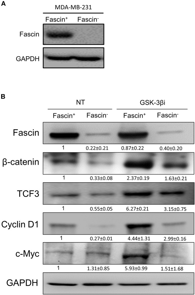
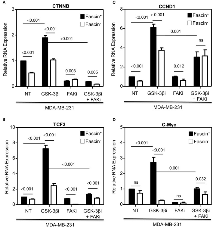
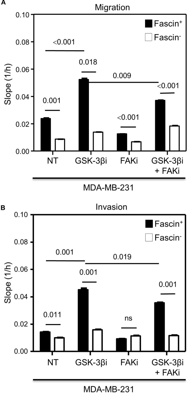
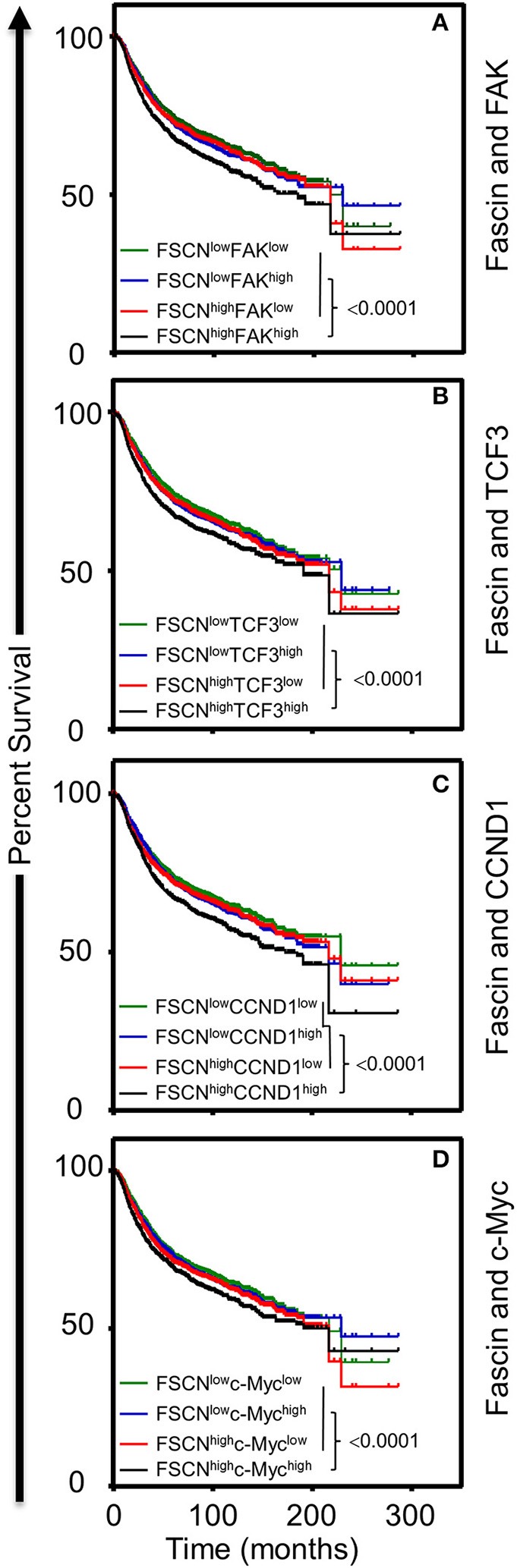
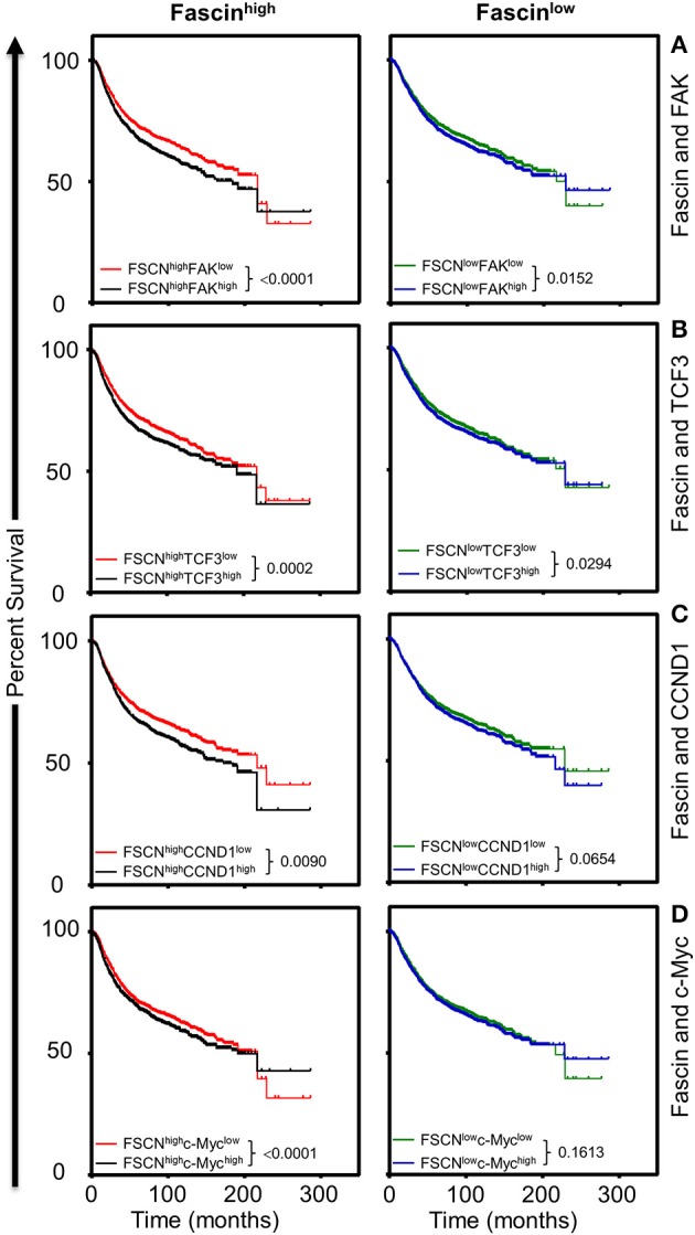
Cancer stem cells (CSCs), a rare population of tumor cells with high self-renewability potential, have gained increasing attention due to their contribution to chemoresistance and metastasis. We have previously demonstrated a critical role for the actin-bundling protein (fascin) in mediating breast cancer chemoresistance through activation of focal adhesion kinase (FAK). The latter is known to trigger the β-catenin signaling pathway. Whether fascin activation of FAK would ultimately trigger β-catenin signaling pathway has not been elucidated. Here, we assessed the effect of fascin manipulation in breast cancer cells on triggering β-catenin downstream targets and its dependence on FAK. Gain and loss of fascin expression showed its direct effect on the constitutive expression of β-catenin downstream targets and enhancement of self-renewability. In addition, fascin was essential for glycogen synthase kinase 3β inhibitor-mediated inducible expression and function of the β-catenin downstream targets. Importantly, fascin-mediated constitutive and inducible expression of β-catenin downstream targets, as well as its subsequent effect on CSC function, was at least partially FAK dependent. To assess the clinical relevance of the in vitro findings, we evaluated the consequence of fascin, FAK, and β-catenin downstream target coexpression on the outcome of breast cancer patient survival. Patients with coexpression of fascinhigh and FAKhigh or high β-catenin downstream targets showed the worst survival outcome, whereas in fascinlow, patient coexpression of FAKhigh or high β-catenin targets had less significant effect on the survival. Altogether, our data demonstrated the critical role of fascin-mediated β-catenin activation and its dependence on intact FAK signaling to promote breast CSC function. These findings suggest that targeting of fascin-FAK-β-catenin axis may provide a novel therapeutic approach for eradication of breast cancer from the root.

Keywords: FAK; breast cancer; cancer stem cell; fascin; β-catenin.


Figures

Figure 1
Figure 1
(A,B) Fascin knockdown reduces the expression of β-catenin and its downstream targets in breast cancer cells. (A) Western blot image showing fascin expression after its knockdown (fascin) in MDA-MB-231 cells. (B) Western blot images showing the expression levels of fascin, β-catenin, TCF3, cyclin D1, and c-Myc following fascin knockdown in MDA-MB-231 cells before and after treatment with GSK-3βi. The expression of each protein was normalized on GAPDH, and the numbers shown below the images indicate the mean fold changes (of three independent experiments ± SD) in fascin in reference to fascin+ group or in GSK-3βi–treated fascin+ and fascin cells in reference to their respective untreated counterparts.
Figure 2
Figure 2
(A–D) Fascin activation of β-catenin downstream target genes is FAK-dependent. Bar graph showing relative RNA expression of CTNNB (A), TCF3 (B), CCND1 (C), and c-Myc (D) following fascin knockdown in MDA-MB-231 cells before and after treatment with GSK-3βi ± FAKi. Results showing the mean of triplicates ± SD of three independent experiments, and each gene is normalized to the expression levels of untreated fascin+ cells.
Figure 3
Figure 3
(A–C) Fascin activation of β-catenin signaling pathway enhances tumorsphere formation in a FAK-dependent manner. Bar graph showing the number of tumorspheres formed by fascin+ and fascin MDA-MB-231 cells before and after treatment with GSK-3βi ± FAKi. (A) Representative images showing primary (top) and secondary (bottom) tumorsphere formed by fascin+ and fascin MDA-MB-231 cells before and after treatment with GSK-3βi ± FAKi. Bar graphs showing primary (B) and secondary (C) tumorspheres as mean of five replicates ± SD of three independent experiments.
Figure 4
Figure 4
(A,B) Fascin activation of β-catenin signaling pathway promotes colony formation in a FAK-dependent manner. (A) Representative images showing colony formation by fascin+ and fascin MDA-MB-231 cells before and after treatment with GSK-3βi ± FAKi. (B) Bar graph showing the number (mean of triplicates ± SD) of colony formed by fascin+ and fascin MDA-MB-231 cells before and after treatment with GSK-3βi ± FAKi. The results are representative of three independent experiments.
Figure 5
Figure 5
(A,B) Fascin activation of β-catenin signaling pathway increases migration and invasion in a FAK-dependent manner. The rate of migration and invasion was monitored in real time using the xCELLigence system. Bar graph showing the migration (A) or invasion (B) rate of fascin+ and fascin MDA-MB-231 cells before and after treatment with GSK-3βi ± FAKi. Migration and invasion rates were measured as increases in the slope (1/h) of the curve during 24 h and are mean of triplicates ± SD of three independent experiments.
Figure 6
Figure 6
(A–D) Coexpression of fascinhigh in conjunction with FAKhigh and high β-catenin downstream targets correlates with poor breast cancer survival. Kaplan–Meier plot showing recurrence-free survival (RFS; n = 3,951) of TCGA breast cancer cohort for (A) fascin and FAK, (B) fascin and TCF3, (C) fascin and CCND1, (D) fascin and c-Myc. Patients were grouped into high or low based on the respective gene expression, and significance between expression groups was calculated using the log-rank test. P-values are indicated on each plot.
Figure 7
Figure 7
(A–D) The influence of FAKhigh and high β-catenin downstream targets on poor survival is more significant in fascinhigh breast cancer patients. Kaplan–Meier plot showing the significance of FAK and β-catenin downstream targets on RFS (n = 3,951) according to fascin expression (fascinhigh and fascinlow). Patients were grouped into fascinhigh (left) and fascinlow (right), and the significance of the respective gene expression in each group was calculated using the log-rank test. P-values are indicated on each plot.

Similar articles

Cited by

References

    1. Li L, Neaves WB. Normal stem cells and cancer stem cells: the niche matters. Cancer Res. (2006) 66:4553–7. 10.1158/0008-5472.CAN-05-3986 - DOI - PubMed
    1. Charafe-Jauffret E, Ginestier C, Iovino F, Wicinski J, Cervera N, Finetti P, et al. . Breast cancer cell lines contain functional cancer stem cells with metastatic capacity and a distinct molecular signature. Cancer Res. (2009) 69:1302–13. 10.1158/0008-5472.CAN-08-2741 - DOI - PMC - PubMed
    1. Yoder BJ, Tso E, Skacel M, Pettay J, Tarr S, Budd T, et al. . The expression of fascin, an actin-bundling motility protein, correlates with hormone receptor-negative breast cancer and a more aggressive clinical course. Clin Cancer Res. (2005) 11:186–92. - PubMed
    1. Al-Alwan M, Olabi S, Ghebeh H, Barhoush E, Tulbah A, Al-Tweigeri T, et al. . Fascin is a key regulator of breast cancer invasion that acts via the modification of metastasis-associated molecules. PLoS ONE. (2011) 6:e27339. 10.1371/journal.pone.0027339 - DOI - PMC - PubMed
    1. Ghebeh H, Al-Khaldi S, Olabi S, Al-Dhfyan A, Al-Mohanna F, Barnawi R, et al. . Fascin is involved in the chemotherapeutic resistance of breast cancer cells predominantly via the PI3K/Akt pathway. Br J Cancer. (2014) 111:1552–61. 10.1038/bjc.2014.453 - DOI - PMC - PubMed