. 2020 Apr 21:10:624.
doi: 10.3389/fonc.2020.00624. eCollection 2020.

Up-Regulation of Activating Transcription Factor 3 in Human Fibroblasts Inhibits Melanoma Cell Growth and Migration Through a Paracrine Pathway

Affiliations

Up-Regulation of Activating Transcription Factor 3 in Human Fibroblasts Inhibits Melanoma Cell Growth and Migration Through a Paracrine Pathway

Tingjian Zu et al. Front Oncol. .

Abstract

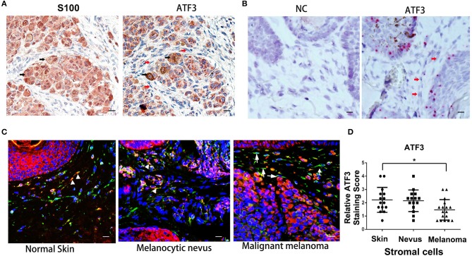
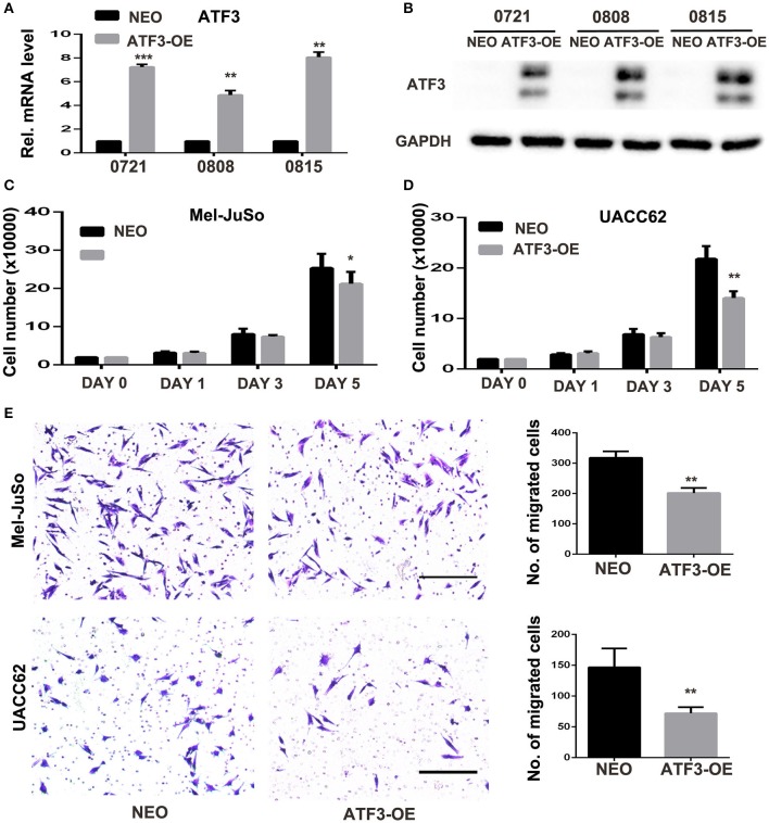
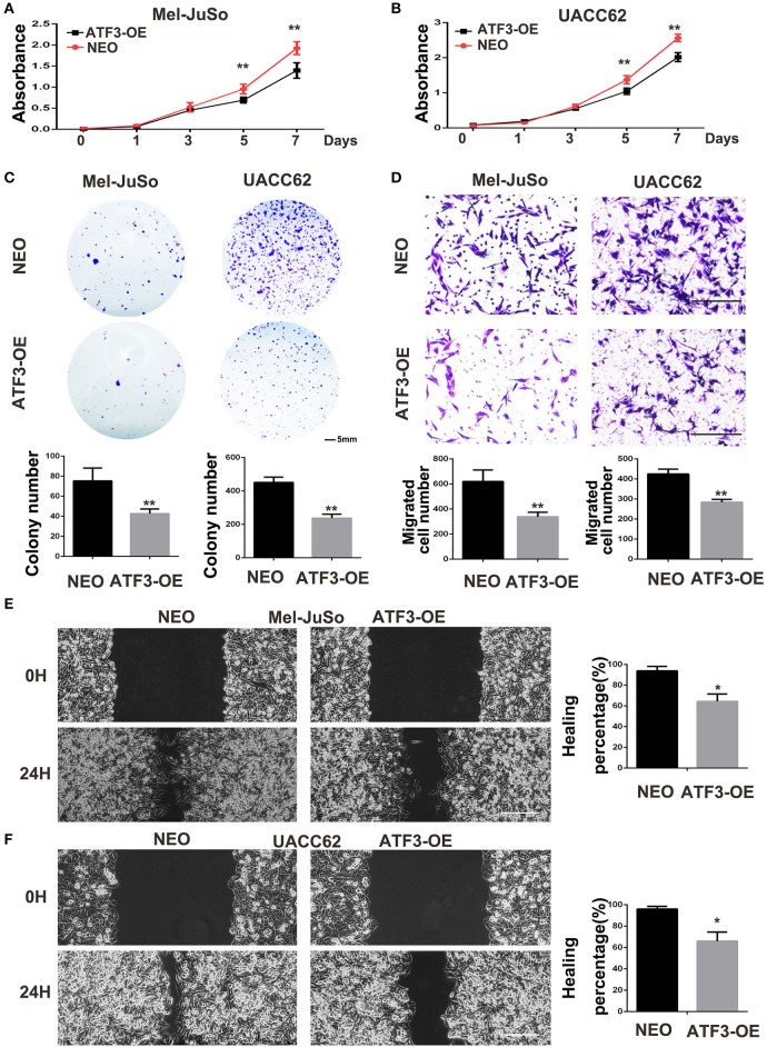
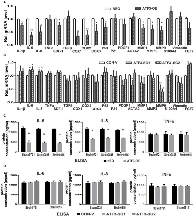
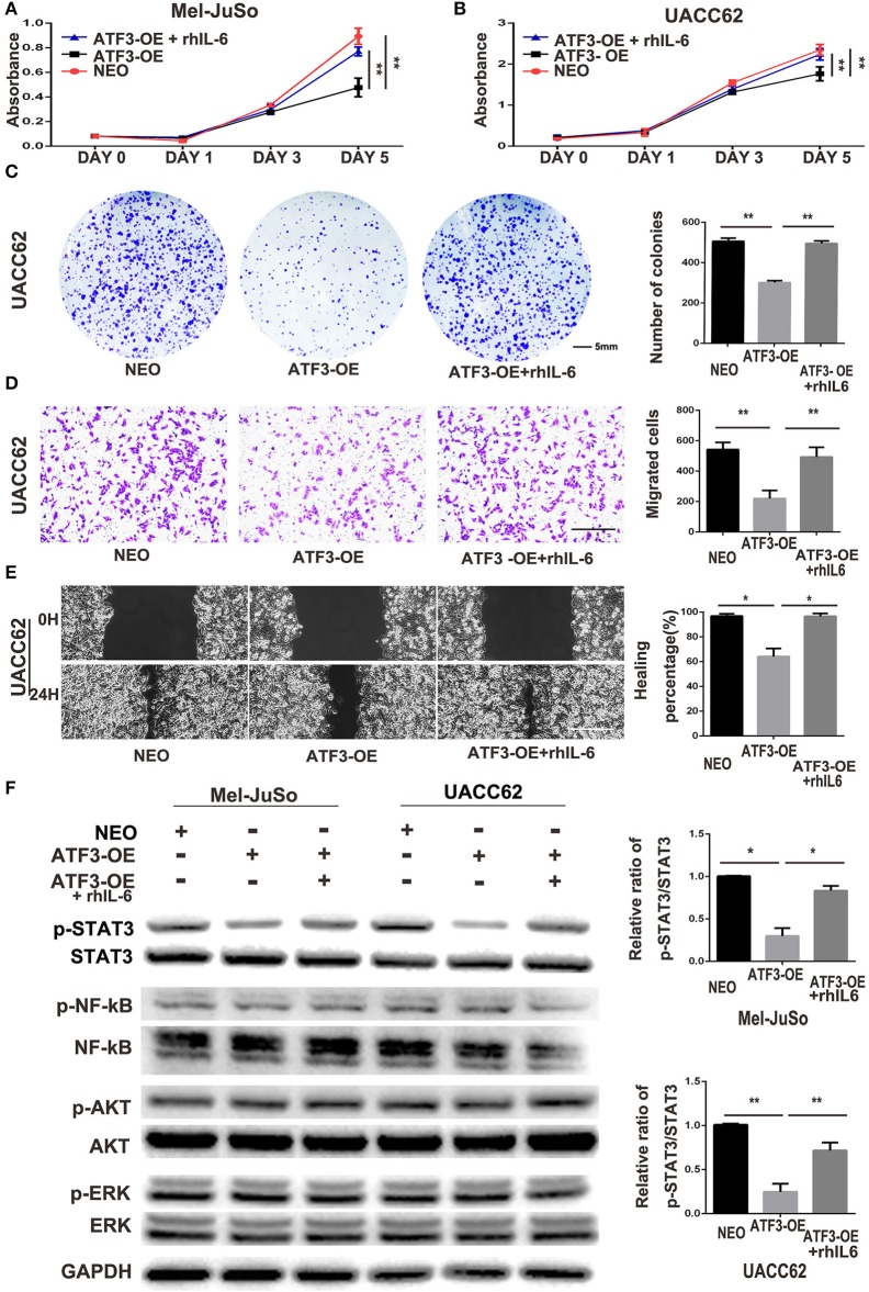
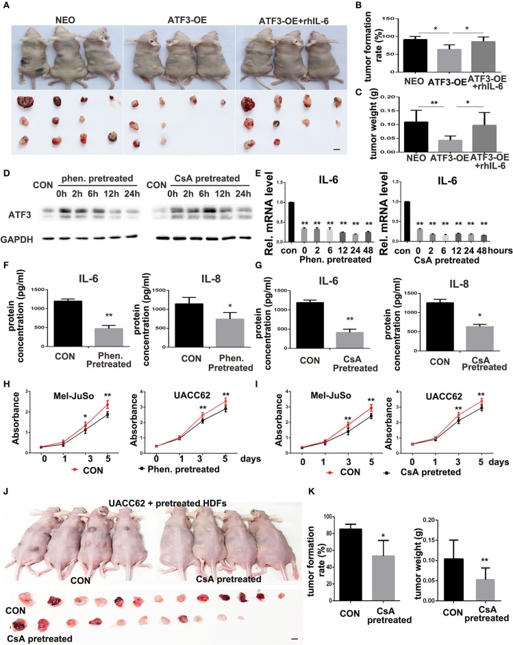
The treatment of melanoma has remained a difficult challenge. Targeting the tumor stroma has recently attracted attention for developing novel strategies for melanoma therapy. Activating transcription factor 3 (ATF3) plays a crucial role in regulating tumorigenesis and development, but whether the expression of ATF3 in human dermal fibroblasts (HDFs) can affect melanoma development hasn't been studied. Our results show that ATF3 expression is downregulated in stromal cells of human melanoma. HDFs expressing high levels of ATF3 suppressed the growth and migration of melanoma cells in association with downregulation of different cytokines including IL-6 in vitro. In vivo, HDFs with high ATF3 expression reduced tumor formation. Adding recombinant IL-6 to melanoma cells reversed those in vitro and in vivo effects, suggesting that ATF3 expression by HDFs regulates melanoma progression through the IL-6/STAT3 pathway. More importantly, HDFs pretreated with cyclosporine A or phenformin to induce ATF3 expression inhibited melanoma cell growth in vitro and in vivo. In summary, our study reveals that ATF3 suppresses human melanoma growth and that inducing the expression of ATF3 in HDFs can inhibit melanoma growth, a new potential melanoma therapeutic approach.

Keywords: activating transcription factor 3; fibroblasts; interleukin 6; melanoma; stromal cells.


Figures

Figure 1
Figure 1
Low expression of ATF3 in human melanoma stromal cells. (A) Immunohistochemistry staining for S100 and ATF3 in clinical melanoma tissues. Black arrows indicate melanoma cells and red arrows indicate stromal cells. (B) RNAscope in situ hybridization staining for ATF3 and negative control (NC) in melanoma samples. Red arrows indicate stromal cells. (C) Double immunofluorescence staining of ATF3 (red) and vimentin (green) in normal skin, melanocytic nevus and melanoma tissues. White arrowheads indicate stromal cells with positive staining of both vimentin (green) and ATF3 (red); white arrows indicate stromal cells with vimentin positive and ATF3 negative staining. (D) Relative scores of ATF3 protein expression levels in stromal cells, detected by vimentin staining as in (C) and quantified nuclear staining of ATF3 from all staining of 15 normal human skin, 15 melanocytic nevus and 17 melanoma tissues. Data are presented as the mean ± standard deviation, *p < 0.05. All scale bars represent 20 μm.
Figure 2
Figure 2
ATF3-overexpressing human dermal fibroblasts (HDFs) inhibited melanoma cell proliferation and migration in co-culture assays. (A) qRT-PCR analysis of ATF3 mRNA levels and (B) Western blot analysis of ATF3 protein with GAPDH as loading control, in three HDF strains infected with ATF3-expressing (ATF3-OE) or control neomycin (NEO)-expressing retrovirus. (C,D) The numbers of Mel-JuSo (C) and UACC62 (D) melanoma cells after co-culture for the indicated number of days with the 3 HDF strains overexpressing ATF3 (ATF3-OE) or expressing NEO. (E) Images (left panels) and quantification (graphs) of Mel-JuSo and UACC62 cells that had migrated after co-culture for 24 h with ATF3-OE or control NEO HDFs. Bars represent 100 μm. Data are presented as the mean ± standard deviation, *p < 0.05, **p < 0.01, ***p < 0.001.
Figure 3
Figure 3
Conditioned medium (CM) derived from ATF3-overexpressing HDFs inhibited melanoma cell growth and migration. CM derived from either ATF3-OE or control NEO HDFs were used for all conditions. (A,B) The proliferation of melanoma cells was analyzed at the indicated times. (C) Colony formation assay was performed; the lower panel graphs show the average number of colonies formed in each group. (D) Melanoma cells that had migrated after 24 h of culture; the average numbers of migrated cells are shown in the lower panel graphs. (E,F) Wound healing assay, images of the cells right after scratching (0H) and 24 h after scratching (24H) are shown in the left panel and percentages of wound closure at 24 h are shown in the right panel graphs. Data are presented as the mean ± standard deviation, *p < 0.05, **p < 0.01. The scale bars represent 5 mm (C) and 100 μm (D,E).
Figure 4
Figure 4
ATF3 overexpression suppressed expression of pro-inflammatory cytokines IL-6 and IL-8 in HDFs. (A) The mRNA levels of cytokine, growth factor and CAF-related genes were measured by qRT-PCR in ATF3-OE HDFs and control NEO HDFs. (B) The mRNA levels of the same genes as in (A) were measured by qRT-PCR in two HDF pools with CRISPR/Cas9-mediated ATF3 deletion by independent small guide RNAs (ATF3-SG1 or ATF3-SG2) and compared with the mRNA levels in control HDFs transduced with empty vector (CON-V). (C,D) Protein concentrations of IL-6, IL-8 and TNFα measured by ELISA in CM derived from ATF3-OE or control NEO HDFs (C) and in CM from HDFs with CRISPR/Cas9-mediated deletion of ATF3 (ATF3-SG1 or ATF3-SG2) or control vector (CON-V) (D). Data are presented as the mean ± standard deviation, *p < 0.05, **p < 0.01.
Figure 5
Figure 5
ATF3-overexpressing HDFs inhibit melanoma cell proliferation and migration through regulation of the IL-6/STAT3 pathway. (A–F) CM derived from either ATF3-OE or control NEO HDFs, or from ATF3-OE HDFs incubated with 10 ng/ml recombinant human IL-6 (ATF3-OE + rhIL-6) were used to culture melanoma cell lines Mel-JuSo and UACC62 for analysis of growth (A,B), colony formation (C), transwell-migration (D), wound healing (E), and phosphorylated (active) states of the indicated signaling proteins (F). Quantification results for C-F assays are shown in the graphs at right. The relative levels of phosphorylated STAT3 in F were normalized to the respective total STAT3 bands. The scale bars in the images represent 5 mm (C) and 100 μm (D,E). Data are presented as the mean ± standard deviation, *p < 0.05, **p < 0.01.
Figure 6
Figure 6
ATF3-overexpressing HDFs inhibit melanoma formation and growth in vivo. (A) Mice and tumors collected at 3 weeks after grafting with the indicated conditions. rhIL-6 (100 ng/mouse/day) was administrated by intraperitoneal injection. (B) The percentage of tumor formation. (C) The average weight of tumors. (D,E) HDFs pretreated with 1.5 mM phenformin (Phen.), 10 μM CsA, or DMSO vehicle (CON) for 24 h were collected at the indicated times for analysis of ATF3, IL-6, and IL-8. (F,G) CM derived from pretreated HDFs was analyzed for IL-6 and IL-8 levels. (H,I) CCK-8 assay after culturing melanoma cells with CM from pretreated HDFs at the indicated time points. (J,K) Mice and tumors collected 3 weeks after grafting as indicated conditions. The percentage of tumor formation and the average tumor weight are shown in (K). The scale bars: 5 mm. Data are presented as the mean ± standard deviation, *p < 0.05, **p < 0.01.

Similar articles

Cited by

References

    1. Aladowicz E, Ferro L, Vitali GC, Venditti E, Fornasari L, Lanfrancone L. Molecular networks in melanoma invasion and metastasis. Fut Oncol. (2013) 9:713–26. 10.2217/fon.13.9 - DOI - PubMed
    1. Aris M, Barrio MM. Combining immunotherapy with oncogene-targeted therapy: a new road for melanoma treatment. Front Immunol. (2015) 6:46. 10.3389/fimmu.2015.00046 - DOI - PMC - PubMed
    1. Zhou L, Yang K, Andl T, Wickett RR, Zhang Y. Perspective of targeting cancer-associated fibroblasts in melanoma. J Cancer. (2015) 6:717–26. 10.7150/jca.10865 - DOI - PMC - PubMed
    1. Soengas MS, Lowe SW. Apoptosis and melanoma chemoresistance. Oncogene. (2003) 22:3138–51. 10.1038/sj.onc.1206454 - DOI - PubMed
    1. Davis EJ, Johnson DB, Sosman JA, Chandra S. Melanoma: what do all the mutations mean? Cancer. (2018) 124:3490–9. 10.1002/cncr.31345 - DOI - PMC - PubMed