Review
. 2020 Apr 15:8:239.
doi: 10.3389/fcell.2020.00239. eCollection 2020.

MitophAging: Mitophagy in Aging and Disease

Affiliations
Review

MitophAging: Mitophagy in Aging and Disease

Daniela Bakula et al. Front Cell Dev Biol. .

Abstract

Maintaining mitochondrial health is emerging as a keystone in aging and associated diseases. The selective degradation of mitochondria by mitophagy is of particular importance in keeping a pristine mitochondrial pool. Indeed, inherited monogenic diseases with defects in mitophagy display complex multisystem pathologies but particularly progressive neurodegeneration. Fortunately, therapies are being developed that target mitophagy allowing new hope for treatments for previously incurable diseases. Herein, we describe mitophagy and associated diseases, coin the term mitophaging and describe new small molecule interventions that target different steps in the mitophagic pathway. Consequently, several age-associated diseases may be treated by targeting mitophagy.

Keywords: aging; autophagy; interventions; mitophaging; mitophagy; monogenic disorders.


Figures

FIGURE 1
FIGURE 1
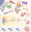
Mitophagy pathways. (A) Ubiquitin-dependent PINK1/Parkin-mediated mitophagy. Upon mitochondrial damage, PINK1 is stabilized at the outer mitochondrial membrane, leading to Parkin activation and subsequent ubiquitination of mitochondrial proteins. Finally, autophagy receptors such as NDP52, OPTN, and p62 are recruited to mediate the engulfment of mitochondria by the autophagosomal membrane through the interaction with LC3. A possible source of the autophagosomal membrane is provided by the endoplasmic reticulum, where the autophagy core complexes VPS34 and ULK1 initiate the membrane formation. The membrane formation is further mediated by WIPI1 and WIPI2, leading to the recruitment of the ATG16L1-complex and LC3, thereby facilitating the formation of autophagosomes. Finally, autophagosomes fuses with acidic lysosomes, a step that is regulated by concerted action of autophagosomal and lysosomal proteins. (B) Ubiquitin-independent receptor-mediated mitophagy. Ubiquitin-independent receptor mediated mitophagy is mediated by the recruitment of autophagy receptor proteins such as NIX, BNIP3, and FUNDC1 to the mitochondrial membrane. The receptor proteins recruit LC3, which enables the engulfment of mitochondria by autophagosomes. (C) Alternative degradation pathways. Piecemeal mitophagy and mitochondrial-derived vesicle degradation are cellular pathways that mediate localized degradation of mitochondria.
FIGURE 2
FIGURE 2
Phenotype clustering of autophagy diseases. (A) Hierarchical clustering of diseases based on the published prevalence of clinical features in the diseases (for data and references see www.mitodb.com). (B) Principal component analysis of diseases based on the prevalence of clinical features. (C) The average prevalence of top-20 clinical features in all autophagy-related disorders (Red, shared with the top-20 features in mitochondrial disorders). (D) The average prevalence of clinical features in mitochondrial diseases.
FIGURE 3
FIGURE 3
Mitophagy interventions. An overview of different mitophagy modulating compounds and their targets. Abbreviations: Ac, Acetylation; HAT, Histone acetyltransferase.

References

    1. Anding A. L., Wang C., Chang T.-K., Sliter D. A., Powers C. M., Hofmann K., et al. (2018). Vps13D encodes a ubiquitin-binding protein that is required for the regulation of mitochondrial size and clearance. Curr. Biol. CB 28:287–295.e6. 10.1016/j.cub.2017.11.064 - DOI - PMC - PubMed
    1. Andreassen S. N., Ben Ezra M., Scheibye-Knudsen M. (2019). A defined human aging phenome. Aging 11 5786–5806. 10.18632/aging.102166 - DOI - PMC - PubMed
    1. Andreux P. A., Blanco-Bose W., Ryu D., Burdet F., Ibberson M., Aebischer P., et al. (2019). The mitophagy activator urolithin A is safe and induces a molecular signature of improved mitochondrial and cellular health in humans. Nat. Metab. 1 595–603. 10.1038/s42255-019-0073-4 - DOI - PubMed
    1. Aparicio R., Rana A., Walker D. W. (2019). Upregulation of the autophagy adaptor p62/SQSTM1 prolongs health and lifespan in middle-aged drosophila. Cell Rep. 28:1029–1040.e5. 10.1016/j.celrep.2019.06.070 - DOI - PMC - PubMed
    1. Bakula D., Müller A. J., Zuleger T., Takacs Z., Franz-Wachtel M., Thost A.-K., et al. (2017). WIPI3 and WIPI4 β-propellers are scaffolds for LKB1-AMPK-TSC signalling circuits in the control of autophagy. Nat. Commun. 8:15637 10.1038/ncomms15637 - DOI - PMC - PubMed

LinkOut - more resources