. 2020 Sep;237(3):556-567.
doi: 10.1111/joa.13203. Epub 2020 May 6.

Disruption of tight junctions contributes to hyposalivation of salivary glands in a mouse model of type 2 diabetes

Affiliations

Disruption of tight junctions contributes to hyposalivation of salivary glands in a mouse model of type 2 diabetes

Yan Huang et al. J Anat. 2020 Sep.

Abstract

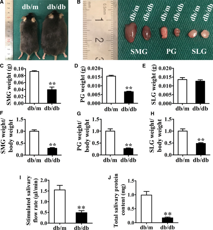
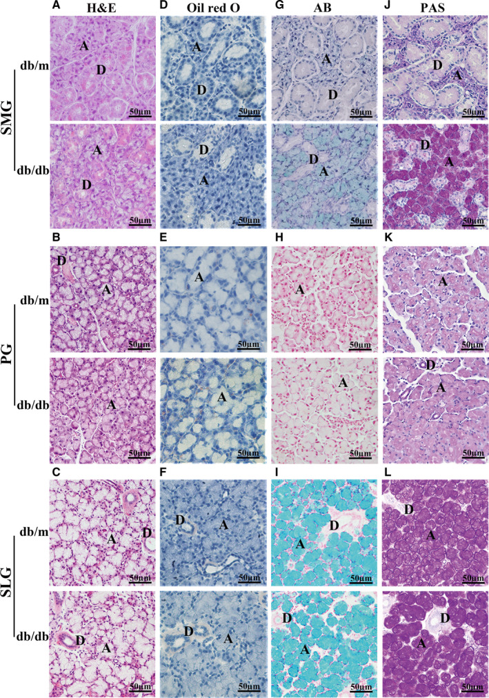
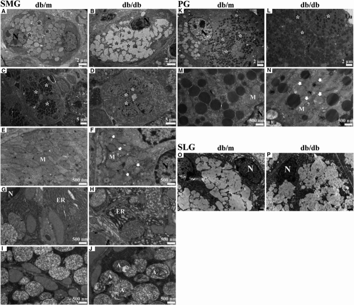
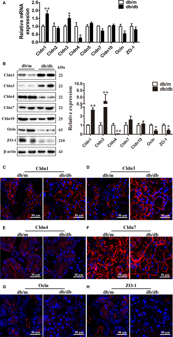
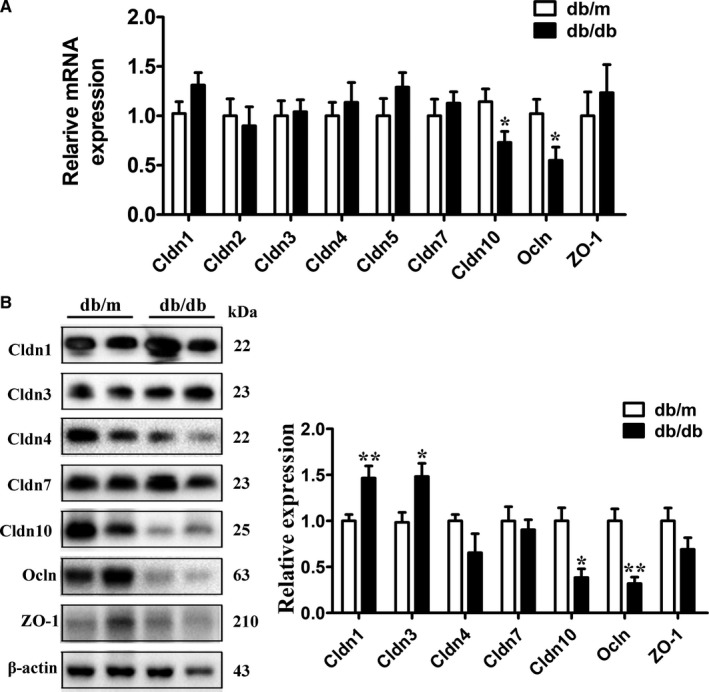
Tight junction (TJ) plays an important role in regulating paracellular fluid transport in salivary glands; however, little is known about the involvement of TJs in diabetes salivary glands. This study aimed to investigate the alterations of TJs and their possible contribution in diabetes-induced hyposalivation. Here, we observed that the morphologies of submandibular glands (SMGs) were impaired, characterized by enlarged acini accumulation with giant secretory granules, which were significantly reduced in atrophic ducts in SMGs of db/db mice, a spontaneous model of type-2 diabetes. However, the secretory granules were increased and scattered in the acini of diabetes parotid glands (PGs). Other ultrastructural damages including swollen mitochondria, expansive endoplasmic reticulum, and autophagosomes were observed in the diabetes group. The levels of TJ proteins including claudin-1 (Cldn1) and claudin-3 (Cldn3) were increased, whereas those of claudin-4 (Cldn4), occludin (Ocln), and zonula occludens-1 (ZO-1) were decreased in SMGs of db/db mice. Higher Cldn1 and Cldn3 and lower claudin-10 (Cldn10) and Ocln levels were observed in PGs of diabetes mice. Taken together, the structures of SMGs and PGs were impaired in diabetes mice, and the disruption of TJ integrity in both SMGs and PGs may contribute to diabetes-induced hyposalivation.

Keywords: diabetes; saliva; salivary gland; submandibular gland; tight junction.


Figures

FIGURE 1
FIGURE 1
Gross morphology and function of submandibular glands, parotid glands and sublingual glands in db/m and db/db mice. (a) Picture of mice. (b) Picture of the three major salivary glands from mice. (c–e) Weight of the glands of SMGs, PGs and SLGs. (f–h) The ratio of gland weight to body weight of SMGs, PGs and SLGs. (i) Stimulated salivary flow rates of db/m and db/db mice. (j) Total salivary protein content from db/m and db/db mice. PG, parotid gland; SLG, sublingual gland; SMG, submandibular gland. Data are presented as mean ± SEM; n = 4–6, **p < .01 compared with db/m mice
FIGURE 2
FIGURE 2
Histological structure of submandibular glands, parotid glands and sublingual glands in db/m and db/db mice. (a–c) H&E staining of SMGs, PGs and SLGs. (d–f) Oil red O staining of SMGs, PGs and SLGs. (g–i) Alcian blue (AB) staining of SMGs, PGs and SLGs. (j–l) Periodic acid‐Schiff (PAS) staining of SMGs, PGs and SLGs. A, acinus; D, duct; PG, parotid gland; SLG, sublingual gland; SMG, submandibular gland
FIGURE 3
FIGURE 3
Ultrastructure of submandibular glands, parotid glands and sublingual glands in db/m and db/db mice. (a, b) Acini of SMGs in db/m and db/db mice. (c, d) Granular convoluted tubules (GCTs) of SMGs in db/m and db/db mice. (e, f) Mitochondria in SMGs of db/m and db/db mice. (g, h) Endoplasmic reticulum in SMGs of db/m and db/db mice. (i, j) Autophagosomes in SMGs of db/m and db/db mice. (k, l) Acini of PGs in db/m and db/db mice. (m, n) Mitochondria in PGs of db/m and db/db mice. (o, p) Acini of SLGs in db/m and db/db mice. A, autophagosomes; ER, endoplasmic reticulum; Lu, lumen; M, mitochondria; N, nucleus; PG, parotid gland; SLG, sublingual gland; SMG, submandibular gland; *, secretory granules; white arrows represent swollen and raptured mitochondria
FIGURE 4
FIGURE 4
Expression of tight junction proteins in submandibular glands of db/m and db/db mice. (a) The mRNA expression of Cldn1, Cldn2, Cldn3, Cldn4, Cldn5, Cldn7, Cldn10, Ocln, and ZO‐1 in SMGs of db/m and db/db mice. (b) The protein levels of Cldn1, Cldn3, Cldn4, Cldn7, Cldn10, Ocln, and ZO‐1 in SMGs of db/m and db/db mice. (c–h) Immunofluorescence staining of Cldn1 (c), Cldn3 (d), Cldn4 (e), Cldn7 (f), Ocln (g), and ZO‐1 (h) in SMGs of db/m and db/db mice. Cldn, claudin; Ocln, occludin; SMG, submandibular gland; ZO‐1, zonula occludens‐1. Data are presented as mean ± SEM; n = 6, *p < .05; **p < .01 compared with db/m mice
FIGURE 5
FIGURE 5
Expression of tight junction proteins in parotid glands of db/m and db/db mice. (a) The mRNA expression of Cldn1, Cldn2, Cldn3, Cldn4, Cldn5, Cldn7, Cldn10, Ocln, and ZO‐1 in PGs of db/m and db/db mice. (b) The protein levels of Cldn1, Cldn3, Cldn4, Cldn7, Cldn10, Ocln, and ZO‐1 in PGs of db/m and db/db mice. PG, parotid gland; Cldn, claudin; Ocln, occludin; ZO‐1, zonula occludens‐1. Data are presented as mean ± SEM; n = 6. *p < .05; **p < .01 compared with db/m mice

Similar articles

Cited by

References

    1. Amoozadeh, Y. , Dan, Q. , Anwer, S. , Huang, H.H. , Barbieri, V. , Waheed, F. et al. (2017) Tumor necrosis factor‐alpha increases claudin‐1, 4, and 7 expression in tubular cells: role in permeability changes. Journal of Cellular Physiology, 232, 2210–2220. - PubMed
    1. Anderson, L.C. and Garrett, J.R. (1986) Lipid accumulation in the major salivary glands of streptozotocin‐diabetic rats. Archives of Oral Biology, 31, 469–475. - PubMed
    1. Anderson, L.C. , Suleiman, A.H. and Garrett, J.R. (1994) Morphological effects of diabetes on the granular ducts and acini of the rat submandibular gland. Microscopy Research and Technique, 27, 61–70. - PubMed
    1. Baker, O.J. (2016) Current trends in salivary gland tight junctions. Tissue Barriers, 4, e1162348. - PMC - PubMed
    1. Beltrame de Oliveira, R. , Matheus, V.A. , Canuto, L.P. , De Sant'ana, A. and Collares‐Buzato, C.B. (2019) Time‐dependent alteration to the tight junction structure of distal intestinal epithelia in type 2 prediabetic mice. Life Sciences, 238, 116971. - PubMed

Publication types

MeSH terms

Substances