. 2020 Aug 15;41(12):3177-3185.
doi: 10.1002/hbm.25006. Epub 2020 May 6.

In vivo characterization of emerging white matter microstructure in the fetal brain in the third trimester

Affiliations

In vivo characterization of emerging white matter microstructure in the fetal brain in the third trimester

Camilo Jaimes et al. Hum Brain Mapp. .

Abstract

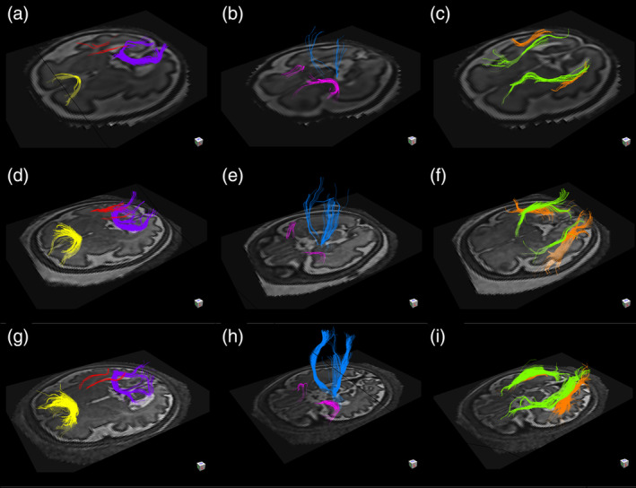
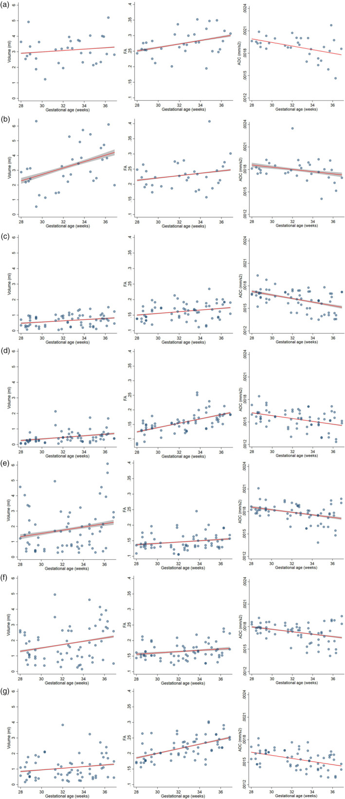
The third trimester of pregnancy is a period of rapid development of fiber bundles in the fetal white matter. Using a recently developed motion-tracked slice-to-volume registration (MT-SVR) method, we aimed to quantify tract-specific developmental changes in apparent diffusion coefficient (ADC), fractional anisotropy (FA), and volume in third trimester healthy fetuses. To this end, we reconstructed diffusion tensor images from motion corrected fetal diffusion magnetic resonance imaging data. With an approved protocol, fetal MRI exams were performed on healthy pregnant women at 3 Tesla and included multiple (2-8) diffusion scans of the fetal head (1-2 b = 0 s/mm2 images and 12 diffusion-sensitized images at b = 500 s/mm2 ). Diffusion data from 32 fetuses (13 females) with median gestational age (GA) of 33 weeks 4 days were processed with MT-SVR and deterministic tractography seeded by regions of interest corresponding to 12 major fiber tracts. Multivariable regression analysis was used to evaluate the association of GA with volume, FA, and ADC for each tract. For all tracts, the volume and FA increased, and the ADC decreased with GA. Associations reached statistical significance for: FA and ADC of the forceps major; volume and ADC for the forceps minor; FA, ADC, and volume for the cingulum; ADC, FA, and volume for the uncinate fasciculi; ADC of the inferior fronto-occipital fasciculi, ADC of the inferior longitudinal fasciculi; and FA and ADC for the corticospinal tracts. These quantitative results demonstrate the complex pattern and rates of tract-specific, GA-related microstructural changes of the developing white matter in human fetal brain.

Keywords: developing white matter; diffusion tensor imaging; diffusion weighted MRI; fetal MRI; fetal brain; tract-specific analysis; tractography; white matter microstructure.


Figures

FIGURE 1
FIGURE 1
Results of in vivo tractography analysis of fetuses at 29 weeks and 3 days (a–c), 32 weeks and 5 days (d–f), and 36 weeks and 1 day (g–i) gestational age. The forceps minor (yellow), the forceps major (purple), and the superior cingulum (red) are depicted in figure parts (a,d,g). The corticospinal tracts (blue) and the uncinate fasciculi (pink) are depicted in figure parts (b,e,h). The inferior longitudinal fasciculi (orange) and the inferior fronto‐occipital fasciculi (green) are depicted in figure parts (c,f,i)
FIGURE 2
FIGURE 2
Changes in volume, FA, and ADC with GA of forceps major (2a) and minor (2b) of the corpus callosum, UF (2c), cingulum (2d), ILF(2e), IFOF (2f) and CSTs (2 g). Fitted linear regression models controlling for sex, motion, and laterality (when applicable) for tract volume, FA, and ADC. 95% confidence intervals of fitted models are shown in gray. ADC, apparent diffusion coefficient; CST, corticospinal tract; FA, fractional anisotropy; IFOF, inferior fronto‐occipital fasciculus, ILF, inferior longitudinal fasciculus; UF, uncinate fasciculus

References

    1. Anastasopoulos, C. , Reisert, M. , Kiselev, V. G. , Nguyen‐Thanh, T. , Schulze‐Bonhage, A. , Zentner, J. , & Mader, I. (2014). Local and global fiber tractography in patients with epilepsy. AJNR. American Journal of Neuroradiology, 35(2), 291–296. 10.3174/ajnr.A3752 - DOI - PMC - PubMed
    1. Barkovich, A. J. , Kjos, B. O. , Jackson, D. E. , & Norman, D. (1988). Normal maturation of the neonatal and infant brain: MR imaging at 1.5 T. Radiology, 166(1 Pt 1), 173–180. 10.1148/radiology.166.1.3336675 - DOI - PubMed
    1. Bouyssi‐Kobar, M. , du Plessis, A. J. , McCarter, R. , Brossard‐Racine, M. , Murnick, J. , Tinkleman, L. , … Limperopoulos, C. (2016). Third trimester brain growth in preterm infants compared with in utero healthy fetuses. Pediatrics, 138(5), e20161640 10.1542/peds.2016-1640 - DOI - PMC - PubMed
    1. Braga, R. M. , Roze, E. , Ball, G. , Merchant, N. , Tusor, N. , Arichi, T. , … Counsell, S. J. (2015). Development of the corticospinal and callosal tracts from extremely premature birth up to 2 years of age. PLoS One, 10(5), e0125681 10.1371/journal.pone.0125681 - DOI - PMC - PubMed
    1. Brody, B. A. , Kinney, H. C. , Kloman, A. S. , & Gilles, F. H. (1987). Sequence of central nervous system myelination in human infancy. I. An autopsy study of myelination. Journal of Neuropathology and Experimental Neurology, 46(3), 283–301. 10.1097/00005072-198705000-00005 - DOI - PubMed

Publication types