Case Reports
. 2020 Nov;77(5):823-831.
doi: 10.1111/his.14138. Epub 2020 Sep 15.

Pulmonary pathology of early-phase COVID-19 pneumonia in a patient with a benign lung lesion

Affiliations
Case Reports

Pulmonary pathology of early-phase COVID-19 pneumonia in a patient with a benign lung lesion

Zhi Zeng et al. Histopathology. 2020 Nov.

Abstract

Aims: An ongoing outbreak of 2019 novel coronavirus (CoV) disease (COVID-19), caused by severe acute respiratory syndrome (SARS) CoV-2, has been spreading in multiple countries. One of the reasons for the rapid spread is that the virus can be transmitted from infected individuals without symptoms. Revealing the pathological features of early-phase COVID-19 pneumonia is important for understanding of its pathogenesis. The aim of this study was to explore the pulmonary pathology of early-phase COVID-19 pneumonia in a patient with a benign lung lesion.

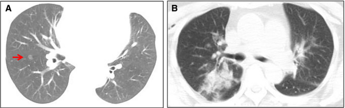
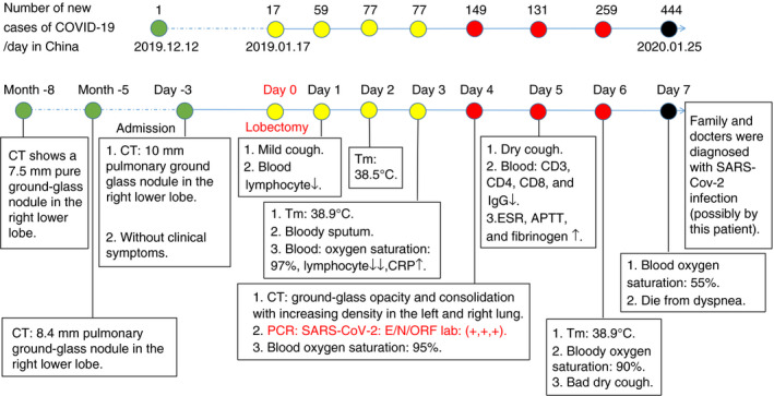
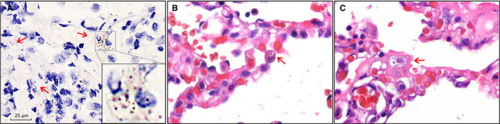
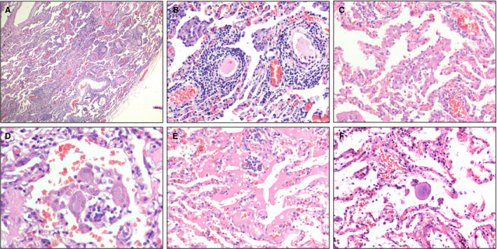
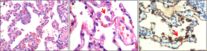
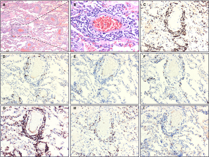
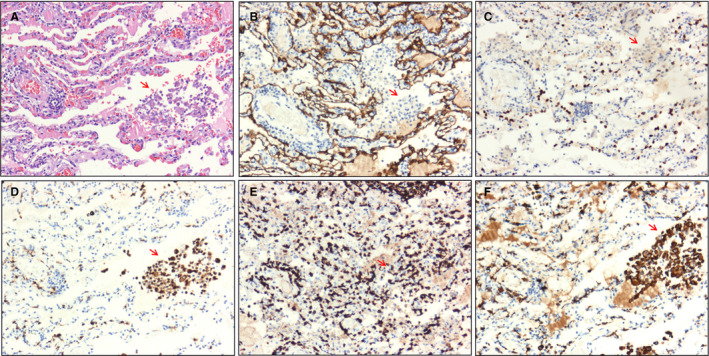
Methods and results: We analysed the pathological changes in lung tissue from a 55-year-old female patient with early-phase SARS-CoV-2 infection. In this case, right lower lobectomy was performed for a benign pulmonary nodule. Detailed clinical, laboratory and radiological data were also examined. This patient was confirmed to have preoperative SARS-CoV-2 infection by the use of real-time reverse transcription polymerase chain reaction and RNA in-situ hybridisation on surgically removed lung tissues. Histologically, COVID-19 pneumonia was characterised by exudative inflammation. The closer to the visceral pleura, the more severe the exudation of monocytes and lymphocytes. Perivascular inflammatory infiltration, intra-alveolar multinucleated giant cells, pneumocyte hyperplasia and intracytoplasmic viral-like inclusion bodies were seen. However, fibrinous exudate and hyaline membrane formation, which were typical pulmonary features of SARS pneumonia, were not evident in this case. Immunohistochemical staining results showed an abnormal accumulation of CD4+ helper T lymphocytes and CD163+ M2 macrophages in the lung tissue.

Conclusion: The results highlighted the pulmonary pathological changes of early-phase SARS-CoV-2 infection, and suggested a role of immune dysfunction in the pathogenesis of COVID-19 pneumonia.

Keywords: 2019 novel coronavirus (SARS-CoV-2); COVID-19 pneumonia; T lymphocyte; macrophage; pathology.


Conflict of interest statement

The authors declare that they have no conflicts of interest.

Figures

Figure 1
Figure 1
Chest computed tomography (CT) images before and after surgery. A, On 14 January 2020, the chest CT image showed a 10‐mm pulmonary ground‐glass nodule (arrow) in the right lower lobe. B, On 21 January 2020, the chest CT image showed patchy ground‐glass opacity and focal consolidation with increasing density in the left and right lungs. [Colour figure can be viewed at wileyonlinelibrary.com]
Figure 2
Figure 2
Clinical history and main laboratory findings. The patient underwent right lower lobectomy on 17 January 2020, and was diagnosed with 2019 novel coronavirus disease (COVID‐19) on postoperative day 4 and died on postoperative day 7. [Colour figure can be viewed at wileyonlinelibrary.com]
Figure 3
Figure 3
Detection of severe acute respiratory syndrome coronavirus 2 (SARS‐CoV‐2) in lung sections. A, SARS‐CoV‐2 RNA was detected with RNA in‐situ hybridisation in lung sections. The positive signals (arrow) were brown in colour and were mainly located in the cytoplasm. Scale bar: 25 μm. Inset: higher magnification of the positive signals. B, An intracytoplasmic viral‐like inclusion body seen in an enlarged epithelial cell (arrow) [haematoxylin and eosin (H&E)]. C, An intracytoplasmic viral‐like inclusion body seen in a macrophage‐like cell (arrow) (H&E).
Figure 4
Figure 4
Pulmonary histomorphology. A, The closer to the visceral pleura, the more severe the exudation of inflammatory cells [haematoxylin and eosin (H&E)]. B, Perivascular inflammatory infiltration (H&E). C, A large number of monocytes, a few lymphocytes and variable numbers of red blood cells were present in the alveolar spaces (H&E). D, Intra‐alveolar multinucleated giant cells (H&E). E, An area of serous exudation (H&E). F, An intra‐alveolar large protein globule (H&E).
Figure 5
Figure 5
Atypical pneumocytes. A, High‐power view of pneumocyte hyperplasia [haematoxylin and eosin (H&E)]. B, High‐power view showing an enlarged pneumocyte (arrow) with amphophilic granular cytoplasm and a prominent eosinophilic nucleolus (H&E). C, Immunohistochemical staining showing atypical pneumocytes to be positive for pan‐cytokeratin (arrow), confirming their epithelial origin (streptavidin peroxidase). [Colour figure can be viewed at wileyonlinelibrary.com]
Figure 6
Figure 6
Immunohistochemical staining of lymphocytes. A, Extensive vasodilation and congestion in lung tissue. The interstitium was widened by oedema and inflammatory cell infiltrates. The alveoli were filled with serous fluid and red blood cells [haematoxylin and eosin (H&E)]. B, Monocytes and lymphocytes around blood vessels (H&E). C, CD3+ T lymphocytes (H&E) and CD20+ and paired box gene 5 (PAX5)+ B lymphocytes. F, Multiple myeloma oncogene‐1 (MUM1)+ plasma cells. G, CD4+ helper T lymphocytes. H, CD8+ cytotoxic T lymphocytes. I, CD56+ natural killer cells (streptavidin peroxidase). [Colour figure can be viewed at wileyonlinelibrary.com]
Figure 7
Figure 7
Immunohistochemical staining of intra‐alveolar cells (arrow). A, Cell clusters consisting of macrophage‐like cells seen in alveolar spaces [haematoxylin and eosin (H&E)]. B,C, Pan‐cytokeratin (PCK)+ and thyroid transcription factor 1 (TTF1)+ alveolar epithelial cells were not detected in intra‐alveolar cell clusters (streptavidin peroxidase). D, CD68+ macrophages were found in alveolar spaces (streptavidin peroxidase). E, CD3+ T lymphocytes were found in alveolar spaces (streptavidin peroxidase). F, Immunohistochemical staining of CD163 showed that most macrophages were of the M2 type (streptavidin peroxidase). [Colour figure can be viewed at wileyonlinelibrary.com]

References

    1. Chan JF, To KK, Tse H, Jin DY, Yuen KY. Interspecies transmission and emergence of novel viruses: lessons from bats and birds. Trends Microbiol. 2013; 21; 544–555. - PMC - PubMed
    1. Summary of probable SARS cases with onset of illness from 1 November 2002 to 31 July 2003. World Health Organization. Available at: https://www.who.int/csr/sars/country/table2004_04_21/en/ (accessed 8 March 2020).
    1. Middle East respiratory syndrome coronavirus (MERS‐CoV). World Health Organization. Available at: http://www.who.int/emergencies/mers-cov/en/ (accessed 8 March 2020).
    1. Update on the novel coronavirus pneumonia outbreak. China National Health Commission. Available at: http://www.nhc.gov.cn/xcs/yqtb/202004/c5c446c763184509ad8baae9f748ab74.s... (accessed 24 April 2020).
    1. Holshue ML, DeBolt C, Lindquist S et al. First case of 2019 novel coronavirus in the United States. N. Engl. J. Med. 2020; 382; 929–936. - PMC - PubMed

Publication types