

Preoperative imaging for colorectal liver metastases: a nationwide population-based study
- PMID: 32374497
- PMCID: PMC7397351
- DOI: 10.1002/bjs5.50291
Preoperative imaging for colorectal liver metastases: a nationwide population-based study
Abstract
Background: In patients with colorectal liver metastases (CRLM) preoperative imaging may include contrast-enhanced (ce) MRI and [18 F]fluorodeoxyglucose (18 F-FDG) PET-CT. This study assessed trends and variation between hospitals and oncological networks in the use of preoperative imaging in the Netherlands.
Methods: Data for all patients who underwent liver resection for CRLM in the Netherlands between 2014 and 2018 were retrieved from a nationwide auditing database. Multivariable logistic regression analysis was used to assess use of ceMRI, 18 F-FDG PET-CT and combined ceMRI and 18 F-FDG PET-CT, and trends in preoperative imaging and hospital and oncological network variation.
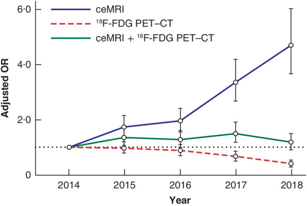
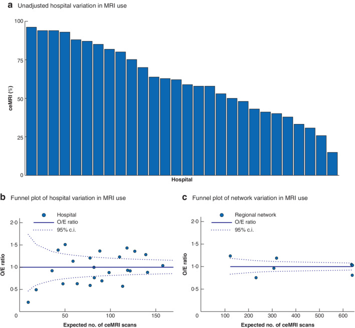
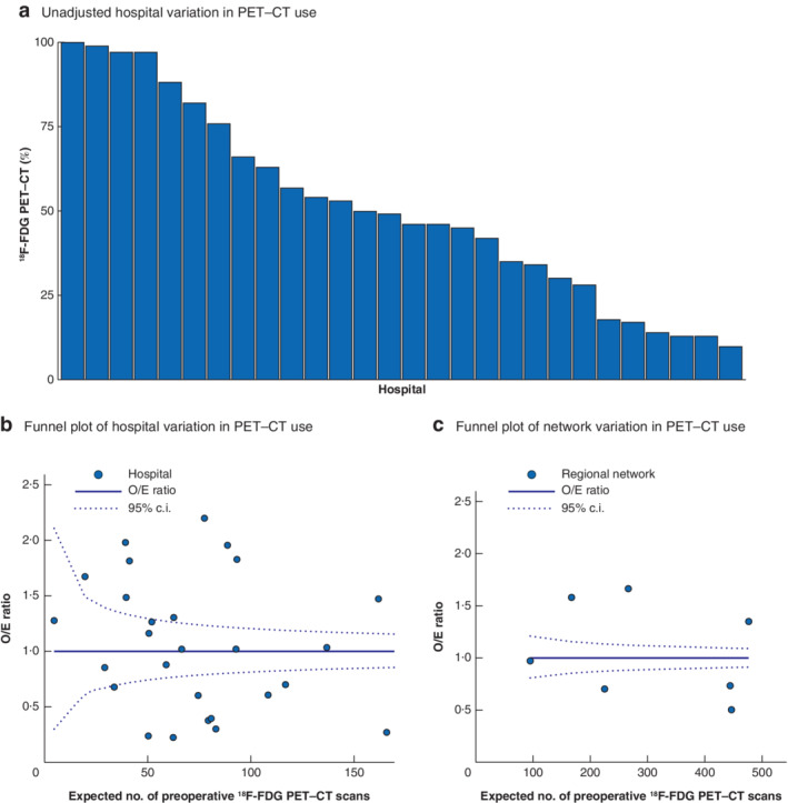
Results: A total of 4510 patients were included, of whom 1562 had ceMRI, 872 had 18 F-FDG PET-CT, and 1293 had combined ceMRI and 18 F-FDG PET-CT. Use of ceMRI increased over time (from 9·6 to 26·2 per cent; P < 0·001), use of 18 F-FDG PET-CT decreased (from 28·6 to 6·0 per cent; P < 0·001), and use of both ceMRI and 18 F-FDG PET-CT 16·9 per cent) remained stable. Unadjusted variation in the use of ceMRI, 18 F-FDG PET-CT, and combined ceMRI and 18 F-FDG PET-CT ranged from 5·6 to 100 per cent between hospitals. After case-mix correction, hospital and oncological network variation was found for all imaging modalities.
Discussion: Significant variation exists concerning the use of preoperative imaging for CRLM between hospitals and oncological networks in the Netherlands. The use of MRI is increasing, whereas that of 18 F-FDG PET-CT is decreasing.
Antecedentes: En pacientes con metástasis hepáticas colorrectales (colorrectal liver metastases, CRLM), los estudios de imagen preoperatorios pueden incluir resonancia magnética con contraste (ce)MRI y 18 F-FDG-PET-CT. Este estudio evaluó las tendencias y la variación entre los hospitales y las redes oncológicas en el uso de estudios de imagen preoperatorios en los Países Bajos. MÉTODOS: Todos los pacientes que se sometieron a una resección hepática por CRLM en los Países Bajos entre 2014 y 2018 fueron seleccionados a partir de una base de datos a nivel nacional auditada. El análisis de regresión logística multivariable se utilizó para evaluar el uso de ceMRI, de 18 F-FDG-PET-CT y de ceMRI combinado con 18 F-FDG-PET-CT, así como para determinar las tendencias en los estudios de imagen preoperatorios y las variaciones hospitalarias y de la red oncológica.
Resultados: En total, se incluyeron 4.510 pacientes, de los cuales 1.562 se sometieron a ceMRI, 872 a 18 F-FDG-PET-CT y 1.293 a ceMRI combinado con 18 F-FDG-PET-CT. El uso de ceMRI aumentó con el tiempo del 9,6% al 26,2% (P < 0,001), el uso de 18 F-FDG-PET-CT disminuyó (25% a 6,0%, P < 0,001) y el uso de ceMRI y 18 F-FDG-PET- CT (17%) se mantuvo estable. La variación no ajustada entre hospitales en el uso de ceMRI, 18 F-FDG-PET-CT y la combinación de ceMRI y 18 F-FDG-PET-CT oscilaba del 5% al 10%. Después de la corrección por case-mix, la variación hospitalaria y de la red oncológica persistía en todas las pruebas de imagen. CONCLUSIÓN: En los Países Bajos existe una variación significativa entre hospitales y redes oncológicas respecto al uso de pruebas de imagen preoperatorias para el CRLM. El uso de MRI está aumentando, mientras que el uso de 18 F-FDG-PET-CT está disminuyendo.
© 2020 The Authors. BJS Open published by John Wiley & Sons Ltd on behalf of British Journal of Surgery Society.
Figures

References
-
- Netherlands Comprehensive Cancer Organization (IKNL). Dutch Cancer Registry; 2018. https://www.iknl.nl/kankersoorten/darmkanker/registratie/incidentie [accessed 27 September 2019].
-
- Takahashi H, Kahramangil B, Kose E, Berber E. A comparison of microwave thermosphere versus radiofrequency thermal ablation in the treatment of colorectal liver metastases. HPB (Oxford) 2018; 20: 1157–1162. - PubMed
-
- Maffione AM, Lopci E, Bluemel C, Giammarile F, Herrmann K, Rubello D. Diagnostic accuracy and impact on management of 18F‐FDG PET and PET/CT in colorectal liver metastasis: a meta‐analysis and systematic review. Eur J Nucl Med Mol Imaging 2015; 42: 152–163. - PubMed
-
- Parikh U, Marcus C, Sarangi R, Taghipour M, Subramaniam RM. FDG PET/CT in pancreatic and hepatobiliary carcinomas: value to patient management and patient outcomes. PET Clin 2015; 10: 327–343. - PubMed
MeSH terms
Substances
LinkOut - more resources
Full Text Sources
Medical
