Meta-Analysis
. 2020 May 6;15(5):e0232856.
doi: 10.1371/journal.pone.0232856. eCollection 2020.

Diagnostic performance of breast tumor tissue selection in diffusion weighted imaging: A systematic review and meta-analysis

Affiliations
Meta-Analysis

Diagnostic performance of breast tumor tissue selection in diffusion weighted imaging: A systematic review and meta-analysis

M Wielema et al. PLoS One. .

Abstract

Background: Several methods for tumor delineation are used in literature on breast diffusion weighted imaging (DWI) to measure the apparent diffusion coefficient (ADC). However, in the process of reaching consensus on breast DWI scanning protocol, image analysis and interpretation, still no standardized optimal breast tumor tissue selection (BTTS) method exists. Therefore, the purpose of this study is to assess the impact of BTTS methods on ADC in the discrimination of benign from malignant breast lesions in DWI in terms of sensitivity, specificity and area under the curve (AUC).

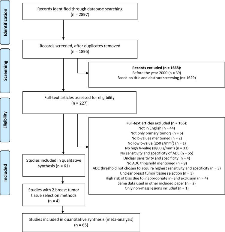
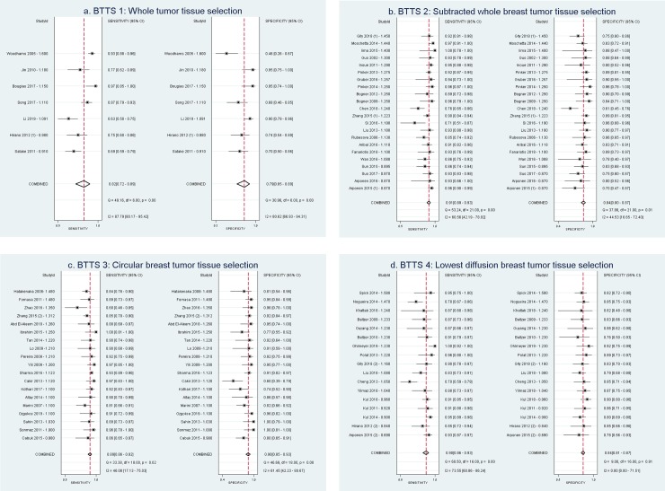
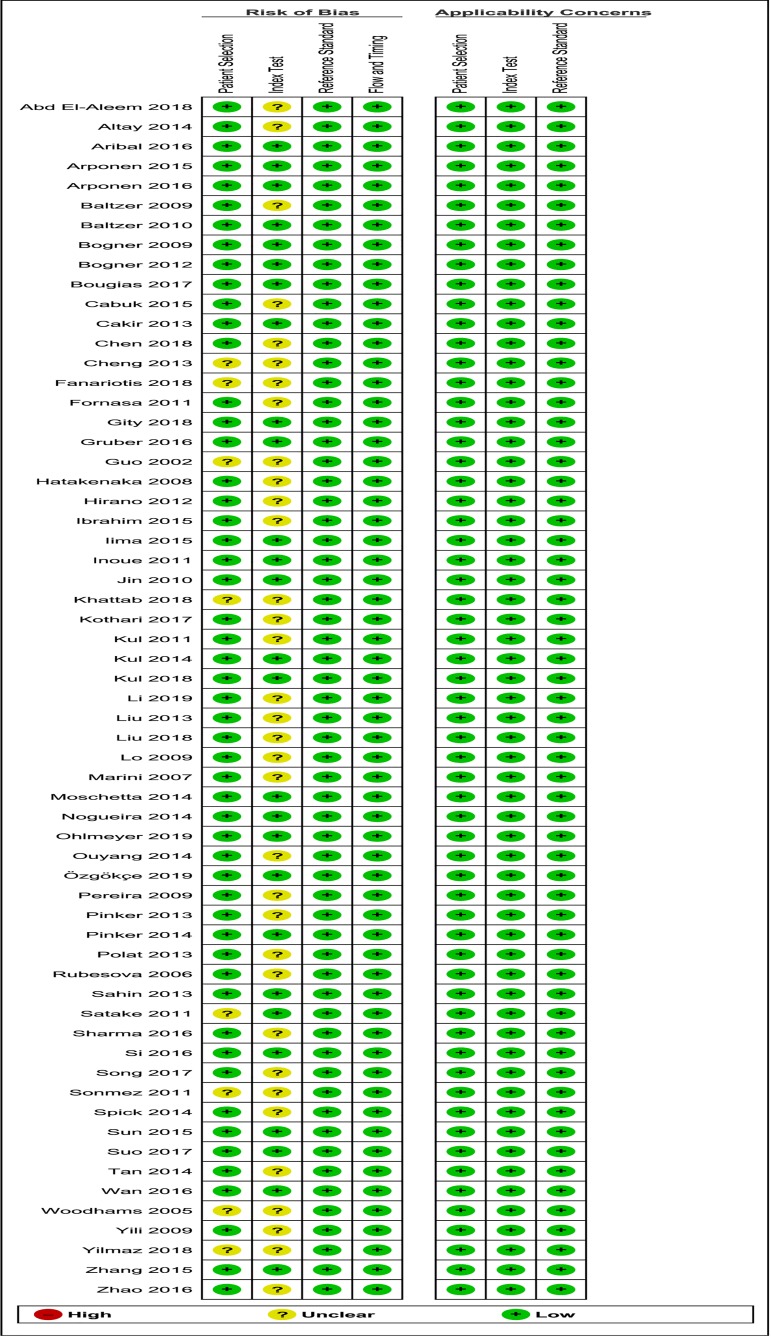
Methods and findings: In this systematic review and meta-analysis, adhering to the PRISMA statement, 61 studies, with 65 study subsets, in females with benign or malignant primary breast lesions (6291 lesions) were assessed. Studies on DWI, quantified by ADC, scanned on 1.5 and 3.0 Tesla and using b-values 0/50 and ≥ 800 s/mm2 were included. PubMed and EMBASE were searched for studies up to 23-10-2019 (n = 2897). Data were pooled based on four BTTS methods (by definition of measured region of interest, ROI): BTTS1: whole breast tumor tissue selection, BTTS2: subtracted whole breast tumor tissue selection, BTTS3: circular breast tumor tissue selection and BTTS4: lowest diffusion breast tumor tissue selection. BTTS methods 2 and 3 excluded necrotic, cystic and hemorrhagic areas. Pooled sensitivity, specificity and AUC of the BTTS methods were calculated. Heterogeneity was explored using the inconsistency index (I2) and considering covariables: field strength, lowest b-value, image of BTTS selection, pre-or post-contrast DWI, slice thickness and ADC threshold. Pooled sensitivity, specificity and AUC were: 0.82 (0.72-0.89), 0.79 (0.65-0.89), 0.88 (0.85-0.90) for BTTS1; 0.91 (0.89-0.93), 0.84 (0.80-0.87), 0.94 (0.91-0.96) for BTTS2; 0.89 (0.86-0.92), 0.90 (0.85-0.93), 0.95 (0.93-0.96) for BTTS3 and 0.90 (0.86-0.93), 0.84 (0.81-0.87), 0.86 (0.82-0.88) for BTTS4, respectively. Significant heterogeneity was found between studies (I2 = 95).

Conclusions: None of the breast tissue selection (BTTS) methodologies outperformed in differentiating benign from malignant breast lesions. The high heterogeneity of ADC data acquisition demands further standardization, such as DWI acquisition parameters and tumor tissue selection to substantially increase the reliability of DWI of the breast.


Conflict of interest statement

The authors have declared that no competing interests exist.

Figures

Fig 1
Fig 1. Flowchart of the study selection process, reasons for excluding papers and numbers of excluded and included papers.
ADC = apparent diffusion coefficient, BTTS = breast tumor tissue selection.
Fig 2
Fig 2. Forest plots for each BTTS method, showing sensitivity and specificity with confidence intervals of each study subset and in the last row the pooled values (diamond).
Study subsets are sorted by ADC threshold. Study ID = Author—ADC threshold (10–3 mm2/s). a) BTTS1: Whole breast tumor tissue selection, b) BTTS2: Subtracted whole breast tumor tissue selection, c) BTTS3: Circular breast tumor tissue selection, d) BTTS4: Lowest diffusion breast tissue selection. BTTS = breast tumor tissue selection. (1)/(2) refers to the two different BTTS method inclusions (study subsets) from the same study.
Fig 3
Fig 3. Summary Receiver Operating Curves of the diagnostic performance of each BTTS method.
SROC with prediction & confidence contours. a) BTTS1: Whole breast tumor tissue selection, b) BTTS2: Subtracted whole breast tumor tissue selection, c) BTTS3: Circular breast tumor tissue selection, d) BTTS4: Lowest diffusion breast tissue selection. BTTS = breast tumor tissue selection, SROC = summarized receiver operator characteristic, AUC = area under the SROC curve, SENS = sensitivity, SPEC = specificity.
Fig 4
Fig 4. Deeks’ funnel plot shows no risk of publication bias.
Fig 5
Fig 5. Summary of the risk of bias and applicability concerns on patient selection, index test, reference standard and flow and timing of the included papers, scored by the reviewers using QUADAS-2 analysis.
QUADAS = Quality Assessment of Diagnostic Accuracy Studies.
Fig 6
Fig 6. Overview of risk of bias and applicability concerns of each included paper, scored by the reviewers using QUADAS-2 analysis.
QUADAS = Quality Assessment of Diagnostic Accuracy Studies.

Similar articles

Cited by

References

    1. Bennani-Baiti B, Bennani-Baiti N, Baltzer PA. Diagnostic performance of breast magnetic resonance imaging in non-calcified equivocal breast findings: Results from a systematic review and meta-analysis. PLoS One. 2016;11: e0160346 10.1371/journal.pone.0160346 - DOI - PMC - PubMed
    1. Dorrius MD, Pijnappel RM, Sijens PE, van der Weide MCJ, Oudkerk M. The negative predictive value of breast Magnetic Resonance Imaging in noncalcified BIRADS 3 lesions. Eur J Radiol. 2012;81: 209–213. 10.1016/j.ejrad.2010.12.046 - DOI - PubMed
    1. Spick C, Pinker-Domenig K, Rudas M, Helbich TH, Baltzer PA, C. S, et al. MRI-only lesions: Application of diffusion-weighted imaging obviates unnecessary MR-guided breast biopsies. Eur Radiol. 2014;24: 1204–1210. 10.1007/s00330-014-3153-6 - DOI - PubMed
    1. Dijkstra H, Dorrius MD, Wielema M, Pijnappel RM, Oudkerk M, Sijens PE. Quantitative DWI implemented after DCE-MRI yields increased specificity for BI-RADS 3 and 4 breast lesions. J Magn Reson Imaging. 2016;44: 1642–1649. 10.1002/jmri.25331 - DOI - PubMed
    1. Baltzer AP, Mann RM, Iima M, Sigmund EE, Clauser P, Gilbert F. Diffusion-Weighted Imaging of the breast–A consensus and mission statement from the EUSOBI International Breast Diffusion- Weighted Imaging working group. Eur Radiol. 2019. - PMC - PubMed