. 2020 Jun 19;48(11):6326-6339.
doi: 10.1093/nar/gkaa324.

Structural basis of the XPB-Bax1 complex as a dynamic helicase-nuclease machinery for DNA repair

Affiliations

Structural basis of the XPB-Bax1 complex as a dynamic helicase-nuclease machinery for DNA repair

Kevin DuPrez et al. Nucleic Acids Res. .

Abstract

Nucleotide excision repair (NER) is a major DNA repair pathway for a variety of DNA lesions. XPB plays a key role in DNA opening at damage sites and coordinating damage incision by nucleases. XPB is conserved from archaea to human. In archaea, XPB is associated with a nuclease Bax1. Here we report crystal structures of XPB in complex with Bax1 from Archaeoglobus fulgidus (Af) and Sulfolobus tokodaii (St). These structures reveal for the first time four domains in Bax1, which interacts with XPB mainly through its N-terminal domain. A Cas2-like domain likely helps to position Bax1 at the forked DNA allowing the nuclease domain to incise one arm of the fork. Bax1 exists in monomer or homodimer but forms a heterodimer exclusively with XPB. StBax1 keeps StXPB in a closed conformation and stimulates ATP hydrolysis by XPB while AfBax1 maintains AfXPB in the open conformation and reduces its ATPase activity. Bax1 contains two distinguished nuclease active sites to presumably incise DNA damage. Our results demonstrate that protein-protein interactions regulate the activities of XPB ATPase and Bax1 nuclease. These structures provide a platform to understand the XPB-nuclease interactions important for the coordination of DNA unwinding and damage incision in eukaryotic NER.


Figures

Figure 1.
Figure 1.
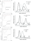
The interactions of XPB with Bax1. (A) The interaction of AfXPB with AfBax1. (B) The interaction of AfXPB-CTD with AfBax1. (C) the interaction of StXPB with StBax1. Left: profiles of ITC titrations; Each titration represents a typical profile of multiple assays with the raw data in the top and data fitting by the ORIGIN software (GE Healthcare) in the bottom. right: profiles of S200 size-exclusion chromatography. The positions of three protein markers are indicated based on chromatographic calibration profile (Supplementary Figure S1) of Conalbumin (75 kDa), Ovalbumin (44 kDa) and Lactalbumin (14 kDa).
Figure 2.
Figure 2.
The two AfXPB–Bax1 heterodimers in the asymmetric unit. (A) Ribbon presentation of the two AfXPB–Bax1 heterodimers. AfXPB molecules are colored in yellow and magenta, AfBax1 molecules are colored in cyan and green. (B) The zoom-in view of the C-termini of AfBax1. (C) Structural comparison of the two AfBax1 molecules. (D) Structural comparison of the two AfXPB molecules. (E) Structural comparison of AfXPB in the heterodimer (yellow) and free of Bax1 (blue, PDB ID 2FWR, chain D) (1).
Figure 3.
Figure 3.
Structure of the AfXPB–Bax1 complex. (A) Diagrams of domain arrangements in AfXPB and AfBax1. Domains are presented as boxes in different colors with labels: DRD (damage recognition domain), HD1 (helicase domain 1), HD2 (helicase domain 2) and ThM (thumb-like) domains of AfXPB are colored in blue, dark green, green, and magenta; NTD, CRD, NUS, and CTD of AfBax1 are colored in cyan, yellow, cyan with red lines for three conserved nuclease motifs, and cyan. (B) The crystal structure of the AfXPB–Bax1 complex in cartoon. Insertion: the interface between AfXPB and Bax1. Residues equivalent to those mutated in ref. (40) are shown as dot spheres with labels. Two acidic residues for the potential N-terminal nuclease active site are indicated by red labels. (C) Superimposition of VRR-Nuc domain (46) (PDB entry: 4QBN, in green ribbons) over the NUS domain of AfBax1. Active site residues are shown in sticks. (D) Overlay of Cas2-forked DNA (PDB entry: 5DQU) (48) on the CRD of AfBax1. Cas2 is colored in wheat and DNA in brown.
Figure 4.
Figure 4.
AfBax1 contains two distinguished nuclease active sites in the N-terminal domain and the nuclease domain. (A) The N-terminal active site plays a role in Bax1-XPB interactions. Mutation D133A/E135A increases the Kd of the AfXPB–Bax1 complex by 10-fold. ITC titration represents a typical profile of multiple assays with the raw data in the top and data fitting by the ORIGIN software (GE Healthcare) in the bottom. (B) DNA incisions on a 16-nt bubble DNA substrate by AfBax1 variants and their complexes with AfXPB. M: DNA oligomer markers, DNA: nuclease reaction control without AfBax1 or AfBax1-XPB complex, wt: wild type AfBax1, n-: AfBax1 mutant D133A/E135A, c-: AfBax1 mutant D305A, n-/c-: AfBax1 mutant D133A/E135A/D305A. Black arrows indicate incised products by the N-terminal nuclease active site (X indicates inhibition on the activity); Grey and light gray arrows indicate incised products by the nuclease domain. (C) Schematic summary of the results from (B). The star indicates P-32 label on the DNA strand.
Figure 5.
Figure 5.
The XPB–Bax1 complex is a dynamic machine. (A) Crystal structure of the StXPB–Bax1 complex in cartoon. StXPB is colored in magenta. StBax1 is colored in blue with CRD in yellow. (B) Superimposition of AfBax1 (in cyan cartoon) with StBax1 (as in A). (C) Superimposition of StXPB from the heterodimer structure (as in A) with the StXPB crystal structure (32) (PDB entry: 5TNU, chain A in gray cartoon) and the AfXPB crystal structure (1) (PDB entry: 2FWR, chain D in green cartoon) over the HD2 and ThM domains. The chain B of StXPB from 5TNU is in the same conformation as StXPB from the heterodimeric structure but is not shown for clear visibility. (D) Structural comparison of the StXPB–Bax1 complex with the AfXPB–Bax1 complex. The AfXPB–Bax1 complex is superimposed with the StXPB–Bax1 complex over the HD2 of AfXPB and StXPB. The AfXPB–Bax1 complex is displayed in ribbons with AfXPB in green and AfBax1 in cyan. Different orientations of the N-terminal half XPB (XPBn) and the C-terminal half Bax1 (Bax1c) between the two heterodimers are highlighted by arrows.
Figure 6.
Figure 6.
The impact of various DNA substrates on the ATPase activity of XPB and the XPB–Bax1 complex. ATPase activities of A. fulgidus (A) or S. tokodaii (B) XPB or the XPB–Bax1 heterodimer were assayed in the absence or presence of a 1:1 molar ratio of protein to DNA for various DNA substrates. ss: single stranded oligonucleotide, 5′-GCCGTGCGCATTCGCCGTGTGGAGCCTGTC-3′; ds: double stranded oligonucleotide, 5′-TGACTCAACATGGAAACCTACAAT-3′; 3′-overhang: -5′-CGAGCACTGCAGTGCTCGTTGTTAT-3′, 3′-GCTCGTGACGTCACGAGC-5′; 5′-overhang: 5′-TATTGTTCGAGCACTGCAGTGCTCG-3′, 3′-GCTCGTGACGTCACGAGC-5′; Y: forked oligonucleotide, 5′-GACAGGCTCACACGTTACGTTGCGCACGGC-3′, 3′-AAAAAAATTCCCGCAATGCAACGCGTGCCG-5′ bubble(6): double stranded oligonucleotide with a 6-bp mismatched bubble in the middle, 5′-TTGACTCAACATCCTTTGCTACAATCAGT-3′, 3′-AACTGAGTTGTATTTCCAGATGTTAGTCA-5′; G4: G-quadruplex oligonucleotide, 5′-TGGACCAGACCTAGCAGCTATGGGGGAGCTGGGGAAGGTGGGAATGTGA-3′; Base-paired nucleotides are underlined. The ATPase activity of StXPB (5.0 μM [Phosphate]/μM protein per minute) and AfXPB (0.4 μM [Phosphate]/μM protein per minute) is used as the base activity in (A) and (B), respectively. The standard deviations are calculated from at least three measurements of the same reaction. (C). AfBax1 hinders the formation of the closed AfXPB conformation. The crystal structure of the AfXPB–Bax1 heterodimer (as in Figure 3A) is superimposed with the closed AfXPB conformation model (1). The N-terminal (DRD and HD1) AfXPB in the closed model is in gray cartoon.
Figure 7.
Figure 7.
Both hydrophobic and charge/polar interactions contribute to the interactions of XPB with Bax1. (A) Biochemical properties of the StXPB surface interacting residues from StBax1: E24, D27, E31, K41, G43, E44, D45, E47, E48, E50YLEKIY56, R62, I83, R86, R87, L89 (label L), F90 (label F), K91YG93, P94 (label P), V95 (label V), L96, E98, R101, I104, I105, M117, V120, F121 and D123LDEE127. Residues selected for mutagenesis are labeled. (B) Biochemical properties of StBax1 surface interacting with residues of StXPB: F278, V282, A285AK287, K289, R292, L295, L296, W298, H299, N303, R316, L319, K323, R332DTQ335, Y338, S341KTFLIPV348, T350YKTD354, E357, E360, I361, K364, E369YRV372, V378 and F379. Residues are represented by sticks and colored in the same way with the partner surface according to amino acid properties: yellow for hydrophobic residues, green for polar uncharged residues, red for acidic residues, and blue for basic residues. Biochemical characteristics of amino acid residues at the Bax1:XPB interface were determined by the PISA server (http://www.ebi.ac.uk/pdbe/prot_int/pistart.html). (C) ITC results for the interaction of StXPB with StBax1 mutant L89A/F90A/P94A/V95A. (D) ITC results for the interaction of StXPB mutant E357A/E360A with StBax1 mutant R86A/R87A. Each ITC titration represents a typical profile of multiple assays with the raw data in the top and data fitting by the ORIGIN software (GE Healthcare) in the bottom.
Figure 8.
Figure 8.
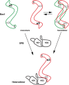
Protein-protein interactions regulate the nuclease activity of Bax1. Bax1 likely contains two nuclease active sites: one (n5) at the N-terminal domain and the other (n3) at the nuclease domain. Bax1 from euryacharchaea A. fulgidus and T. acidophilum is predominantly monomer in solution and is in the conformation preferable for the N-terminal (n5) activity, which presumably incises DNA 5′ to the damage in the middle of the bubble. StBax1 and SsBax1 from crenarchaea Sulfurisphaera are exclusively homodimers in solution and have no apparent nuclease activity (39) because the two nuclease active sites are mutually masked due to dimerization. Both monomeric and dimeric Bax1 interact with XPB to form the heterodimeric XPB–Bax1 complex which masks the N-terminal active site (n5) but enhances the activity of the nuclease domain (n3). The nuclease domain presumably incises DNA 3′ to the damage in the middle of the bubble. Active nuclease sites are highlighted by black labels (n3 or n5) while inhibited nuclease sites are labeled in gray. The C-terminus of Bax1 is indicated by letter C while the two helicase domains of XPB are labeled by HD1 and HD2.

Similar articles

Cited by

References

    1. Fan L., Arvai A.S., Cooper P.K., Iwai S., Hanaoka F., Tainer J.A.. Conserved XPB core structure and motifs for DNA unwinding: implications for pathway selection of transcription or excision repair. Mol. Cell. 2006; 22:27–37. - PubMed
    1. Ma X., Hong Y., Han W., Sheng D., Ni J., Hou G., Shen Y.. Single-stranded DNA binding activity of XPBI, but not XPBII, from Sulfolobus tokodaii causes double-stranded DNA melting. Extremophiles. 2011; 15:67–76. - PubMed
    1. Gulyas K.D., Donahue T.F.. SSL2, a suppressor of a stem-loop mutation in the HIS4 leader encodes the yeast homolog of human ERCC-3. Cell. 1992; 69:1031–1042. - PubMed
    1. Park E., Guzder S.N., Koken M.H., Jaspers-Dekker I., Weeda G., Hoeijmakers J.H., Prakash S., Prakash L.. RAD25 (SSL2), the yeast homolog of the human xeroderma pigmentosum group B DNA repair gene, is essential for viability. Proc. Natl. Acad. Sci. U.S.A. 1992; 89:11416–11420. - PMC - PubMed
    1. Koken M.H., Vreeken C., Bol S.A., Cheng N.C., Jaspers-Dekker I., Hoeijmakers J.H., Eeken J.C., Weeda G., Pastink A.. Cloning and characterization of the Drosophila homolog of the xeroderma pigmentosum complementation-group B correcting gene, ERCC3. Nucleic Acids Res. 1992; 20:5541–5548. - PMC - PubMed

Publication types

MeSH terms